 Explore
 Validate
 Learn
Learn Western blot
 Immunoprecipitation
Antibody data
- Antibody Data
- Antigen structure
- References [0]
- Comments [0]
- Validations
- Western blot [1]
- Immunohistochemistry [3]
Submit
Validation data
Reference
Comment
Report error
- Product number
- PA5-72903 - Provider product page

- Provider
- Invitrogen Antibodies
- Product name
- Caspase 14 Polyclonal Antibody
- Antibody type
- Polyclonal
- Antigen
- Recombinant full-length protein
- Reactivity
- Human, Mouse, Rat, Canine
- Host
- Rabbit
- Isotype
- IgG
- Vial size
- 50 µL
- Concentration
- Conc. Not Determined
- Storage
- Store at 4°C short term. For long term storage, store at -20°C, avoiding freeze/thaw cycles.
No comments: Submit comment
Supportive validation
- Submitted by
- Invitrogen Antibodies (provider)
- Main image
- Experimental details
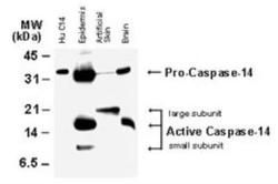
- Western blot analysis of Caspase 14 in tissue lysates (50 µg/lane) and recombinant human Caspase-14 (Hu C14, 15 ng). Samples were incubated in Caspase 14 polyclonal antibody (Product # PA5-72903 using a dilution of 1:2000. The antibody detected both the proform of caspase-14, and the large and small subunits of active/cleaved caspase-14.

Supportive validation
- Submitted by
- Invitrogen Antibodies (provider)
- Main image

- Experimental details
- Immunohistochemical analysis of Caspase 14 in Tissue sections of mouse skin. Samples were incubated in Caspase 14 polyclonal antibody (Product # PA5-72903) using a dilution of 1:500.

- Submitted by
- Invitrogen Antibodies (provider)
- Main image

- Experimental details
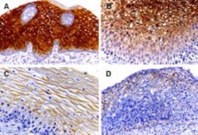
- Immunohistochemical analysis of Caspase 14 in Tissue sections of human cervix. Samples were incubated in Caspase 14 polyclonal antibody (Product # PA5-72903) using a dilution of 1:2000. A. Normal cervix (squamous epithelium). B. CIN1 (low-grade squamous intraepithelial lesion, mild dysplasia). C. CIN2 (high-grade squamous intraepithelial lesion, moderate dysplasia. D. CIN3 (high-grade squamous intraepithelial lesion; severe dysplasia-carcinoma in situ. In normal cervi, caspase-14 staining was found most in the midzone layer, but was absent from the basal/parabasal cell layer where mitotically active cells are known to reside. This suggests induction of caspase-14 expression with differentiation. Caspase-14 expression declined progressively during malignant transformation as the histologic severity of the cervical atypia advanced from CIN1 to CIN3. Hematoxylin-eosin counterstain.
- Submitted by
- Invitrogen Antibodies (provider)
- Main image

- Experimental details
- Immunohistochemical analysis of Caspase 14 in Human ovarian cancer tissue microarray. Samples were incubated in Caspase 14 polyclonal antibody (Product # PA5-72903) using a dilution of 1:2000. Low (A) and high (B) stage ovarian tumor tissue cores. High magnification from areas of the tissue cores (A1 and B1). Decreased Caspase-14 expression was seen in the high grade, compared to the low grade tumor. Hematoxylin-eosin counterstain.
