Explore
 Validate
 Learn
Learn Western blot
 Immunocytochemistry
Antibody data
- Antibody Data
- Antigen structure
- References [0]
- Comments [0]
- Validations
- Immunocytochemistry [6]
Submit
Validation data
Reference
Comment
Report error
- Product number
- OSV00035G-500UG - Provider product page

- Provider
- Invitrogen Antibodies
- Product name
- VDP Polyclonal Antibody
- Antibody type
- Polyclonal
- Antigen
- Synthetic peptide
- Reactivity
- Human, Mouse, Rat
- Host
- Rabbit
- Isotype
- IgG
- Vial size
- 500 µg
- Concentration
- Conc. Not Determined
- Storage
- Store at 4°C short term. For long term storage, store at -20°C, avoiding freeze/thaw cycles. Glycerol (1:1) may be added for added stability.
No comments: Submit comment
Supportive validation
- Submitted by
- Invitrogen Antibodies (provider)
- Main image

- Experimental details
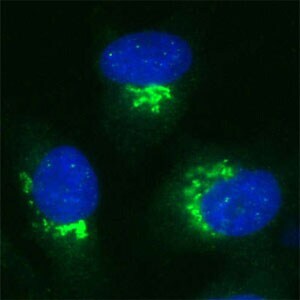
- Human Melanoma cell line C32 was cultured overnight on round cover slides placed in a 24 well tissue culture plate. Culture media removed and washed twice with PBS before fixing with 2% formalin for 10 minutes. Cells were then washed three times with PBS and incubated with Tris 0.01M containing Triton X 0.005% for 15 minutes. Cells were washed and incubated with 100 µL of Rabbit antibody to internal region of Vesicle docking protein (VDP, USO1, TAP, Transcytosis-associated protein): IgG (OSV00035G) diluted 1:100 in the blocking buffer for 30 minutes. Welles were then washed 7 times with PBS and incubated with 100 µL of anti Rb-FITC conjugate diluted 1:100 in the blocking buffer for further 30 minutes. Cells were washed as before and nuclear counter stained with Hoechst and mounted on to slides.
- Submitted by
- Invitrogen Antibodies (provider)
- Main image

- Experimental details
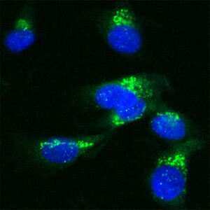
- Human Melanoma cell line C32 was cultured overnight on round cover slides placed in a 24 well tissue culture plate. Culture media removed and washed twice with PBS before fixing with 2% formalin for 10 minutes. Cells were then washed three times with PBS and incubated with Tris 0.01M containing Triton X 0.005% for 15 minutes. Cells were washed and incubated with 100 µL of Rabbit antibody to internal region of Vesicle docking protein (VDP, USO1, TAP, Transcytosis-associated protein): IgG (OSV00035G) diluted 1:100 in the blocking buffer for 30 minutes. Welles were then washed 7 times with PBS and incubated with 100 µL of anti Rb-FITC conjugate diluted 1:100 in the blocking buffer for further 30 minutes. Cells were washed as before and nuclear counter stained with Hoechst and mounted on to slides.
- Submitted by
- Invitrogen Antibodies (provider)
- Main image
- Experimental details
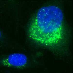
- Human Melanoma cell line C32 was cultured overnight on round cover slides placed in a 24 well tissue culture plate. Culture media removed and washed twice with PBS before fixing with 2% formalin for 10 minutes. Cells were then washed three times with PBS and incubated with Tris 0.01M containing Triton X 0.005% for 15 minutes. Cells were washed and incubated with 100 µL of Rabbit antibody to internal region of Vesicle docking protein (VDP, USO1, TAP, Transcytosis-associated protein): IgG (OSV00035G) diluted 1:100 in the blocking buffer for 30 minutes. Welles were then washed 7 times with PBS and incubated with 100 µL of anti Rb-FITC conjugate diluted 1:100 in the blocking buffer for further 30 minutes. Cells were washed as before and nuclear counter stained with Hoechst and mounted on to slides.

- Submitted by
- Invitrogen Antibodies (provider)
- Main image

- Experimental details
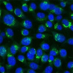
- Human Melanoma cell line C32 was cultured overnight on round cover slides placed in a 24 well tissue culture plate. Culture media removed and washed twice with PBS before fixing with 2% formalin for 10 minutes. Cells were then washed three times with PBS and incubated with Tris 0.01M containing Triton X 0.005% for 15 minutes. Cells were washed and incubated with 100 µL of Rabbit antibody to internal region of Vesicle docking protein (VDP, USO1, TAP, Transcytosis-associated protein): IgG (OSV00035G) diluted 1:100 in the blocking buffer for 30 minutes. Welles were then washed 7 times with PBS and incubated with 100 µL of anti Rb-FITC conjugate diluted 1:100 in the blocking buffer for further 30 minutes. Cells were washed as before and nuclear counter stained with Hoechst and mounted on to slides.

- Submitted by
- Invitrogen Antibodies (provider)
- Main image

- Experimental details
- Human Melanoma cell line C32 was cultured overnight on round cover slides placed in a 24 well tissue culture plate. Culture media removed and washed twice with PBS before fixing with 2% formalin for 10 minutes. Cells were then washed three times with PBS and incubated with Tris 0.01M containing Triton X 0.005% for 15 minutes. Cells were washed and incubated with 100 µL of Rabbit antibody to internal region of Vesicle docking protein (VDP, USO1, TAP, Transcytosis-associated protein): IgG (OSV00035G) diluted 1:100 in the blocking buffer for 30 minutes. Welles were then washed 7 times with PBS and incubated with 100 µL of anti Rb-FITC conjugate diluted 1:100 in the blocking buffer for further 30 minutes. Cells were washed as before and nuclear counter stained with Hoechst and mounted on to slides.