Explore
 Validate
 Learn
Learn Western blot
 Immunocytochemistry
Antibody data
- Antibody Data
- Antigen structure
- References [1]
- Comments [0]
- Validations
- Immunocytochemistry [3]
- Immunohistochemistry [1]
- Other assay [1]
Submit
Validation data
Reference
Comment
Report error
- Product number
- PA5-30281 - Provider product page

- Provider
- Invitrogen Antibodies
- Product name
- VDP Polyclonal Antibody
- Antibody type
- Polyclonal
- Antigen
- Recombinant full-length protein
- Description
- Recommended positive controls: 293T, A431, HeLa, HepG2, NIH-3T3, mouse liver. Predicted reactivity: Human (99%), Mouse (94%), Rat (93%), Chicken (88%), Bovine (99%). Store product as a concentrated solution. Centrifuge briefly prior to opening the vial.
- Reactivity
- Human, Mouse
- Host
- Rabbit
- Isotype
- IgG
- Vial size
- 100 μL
- Concentration
- 0.5 mg/mL
- Storage
- Store at 4°C short term. For long term storage, store at -20°C, avoiding freeze/thaw cycles.
Submitted references Autophagy Inhibition Induces the Secretion of Macrophage Migration Inhibitory Factor (MIF) with Autocrine and Paracrine Effects on the Promotion of Malignancy in Breast Cancer.
Cotzomi-Ortega I, Rosas-Cruz A, Ramírez-Ramírez D, Reyes-Leyva J, Rodriguez-Sosa M, Aguilar-Alonso P, Maycotte P
Biology 2020 Jan 18;9(1)
Biology 2020 Jan 18;9(1)
No comments: Submit comment
Supportive validation
- Submitted by
- Invitrogen Antibodies (provider)
- Main image

- Experimental details
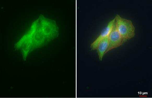
- VDP Polyclonal Antibody [N1N2], N-term detects VDP / p115 protein at cytoplasm and golgi apparatus by immunofluorescent analysis. Sample: HepG2 cells were fixed in ice-cold MeOH for 5 min. Green: VDP / p115 stained by VDP Polyclonal Antibody [N1N2], N-term (Product # PA5-30281) diluted at 1:500. Red: alpha Tubulin, a cytoskeleton marker, stained by alpha Tubulin Polyclonal Antibody [GT114] (Product # MA5-31466) diluted at 1:1,000. Blue: Fluoroshield with DAPI .
- Submitted by
- Invitrogen Antibodies (provider)
- Main image

- Experimental details
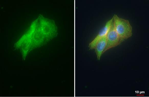
- VDP Polyclonal Antibody [N1N2], N-term detects VDP / p115 protein at cytoplasm and golgi apparatus by immunofluorescent analysis. Sample: HepG2 cells were fixed in ice-cold MeOH for 5 min. Green: VDP / p115 stained by VDP Polyclonal Antibody [N1N2], N-term (Product # PA5-30281) diluted at 1:500. Red: alpha Tubulin, a cytoskeleton marker, stained by alpha Tubulin Polyclonal Antibody [GT114] (Product # MA5-31466) diluted at 1:1,000. Blue: Fluoroshield with DAPI .
- Submitted by
- Invitrogen Antibodies (provider)
- Main image

- Experimental details
- VDP Polyclonal Antibody [N1N2], N-term detects VDP / p115 protein at cytoplasm and golgi apparatus by immunofluorescent analysis. Sample: HepG2 cells were fixed in ice-cold MeOH for 5 min. Green: VDP / p115 stained by VDP Polyclonal Antibody [N1N2], N-term (Product # PA5-30281) diluted at 1:500. Red: alpha Tubulin, a cytoskeleton marker, stained by alpha Tubulin Polyclonal Antibody [GT114] (Product # MA5-31466) diluted at 1:1,000. Blue: Fluoroshield with DAPI .

Supportive validation
- Submitted by
- Invitrogen Antibodies (provider)
- Main image
- Experimental details
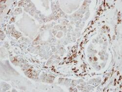
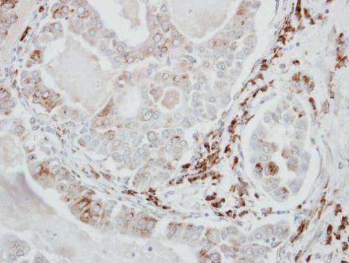
- Immunohistochemical analysis of paraffin-embedded human lung adenocarcinoma, using VDP/p115 (Product # PA5-30281) antibody at 1:500 dilution. Antigen Retrieval: EDTA based buffer, pH 8.0, 15 min.
Supportive validation
- Submitted by
- Invitrogen Antibodies (provider)
- Main image
- Experimental details
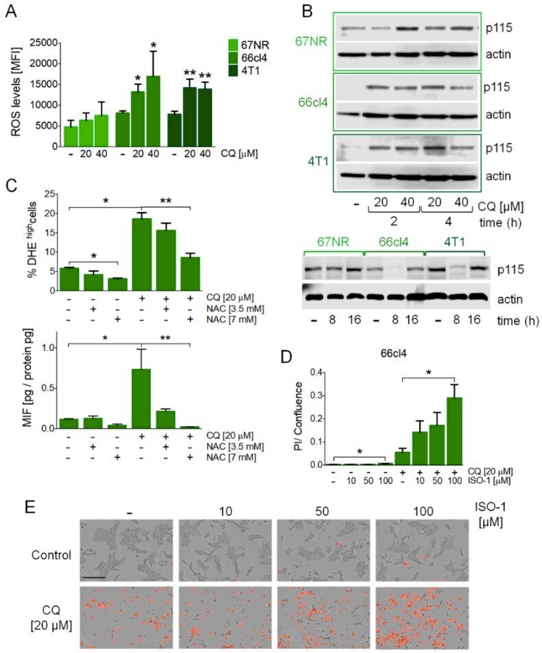
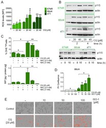
- Figure 3 MIF secretion induced by the inhibition of autophagy was mediated by reactive oxygen species (ROS) production, occurred with changes in Uso1/p115 protein levels, and mediated cell survival in the 66cl4 cell line. ROS levels were evaluated by DHE staining by flow cytometry at 16 h of treatment at the indicated concentrations of CQ ( A ). Uso1/p115 protein levels were evaluated by Western blot in the three cell lines studied ( B ). N-acetyl cysteine (NAC) treatment at the indicated concentrations reduced the ROS levels induced by CQ treatment and reduced MIF secretion in the 66cl4 cell line ( C ) both at 16 h of treatment. ISO-1, a MIF inhibitor, increased cell death in the 66cl4 cell line when used in combination with CQ treatment for 24 h ( D , E ). The scale bar in the pictures in ( E ) represents 200 um. Results show mean +/- standard error of three-four independent experiments, * p < 0.05, ** p < 0.01.