 Explore
 Validate
 Learn
Learn Flow cytometry
Antibody data
- Antibody Data
- Antigen structure
- References [2]
- Comments [0]
- Validations
- Flow cytometry [1]
- Other assay [3]
Submit
Validation data
Reference
Comment
Report error
- Product number
- 64-1278-42 - Provider product page

- Provider
- Invitrogen Antibodies
- Product name
- CD127 Monoclonal Antibody (eBioRDR5), Super Bright™ 645, eBioscience™
- Antibody type
- Monoclonal
- Antigen
- Other
- Description
- Description: The eBioRDR5 monoclonal antibody reacts with human CD127 (Interleukin-7 Receptor alpha). CD127 complexes with CD132, also known as the common gamma chain (gamma c), to form the multi-functional IL-7 receptor (IL-7R). CD127 is a type I glycoprotein with a molecular weight of 75-80 kDa and is expressed by immature B cells through the early pre-B stage, by thymocytes during several stages of their development, and on most mature T cells, with transient down-regulation upon activation. Binding of IL-7 results in signal transduction which occurs through several tyrosine kinase pathways including the Jak/STAT pathway. IL-7 is indispensible for the development of lymphocytes, and the control of homeostatic proliferation of T-cells in the periphery. In addition, IL-7R signaling is know to be involved in the regulation of T cell receptor (TCR) locus rearrangement in gamma delta T cells. Interestingly, recently it has been demonstrated that CD127 expression is down-regulated on CD4+CD25+ regulatory T cells (T regs). While the co-expression of CD4 and CD25 has become widely used as an indicator of T regs, this method of identification may also include cells without suppressive activity. It has clearly been shown that CD4+CD25+ cells that have down-regulated the expression of CD127 are significantly more highly-enriched for the regulatory T population, as defined by expression of the T reg-specific transcription factor Foxp3 and the suppressive activity of these cells, in vitro. Binding of the eBioRDR5 monoclonal antibody to PBMCs is blocked by pre-incubation of the cells with recombinant human IL-7 (Product # 14-1079-80). Applications Reported: This eBioRDR5 antibody has been reported for use in flow cytometric analysis. Applications Tested: This eBioRDR5 antibody has been pre-titrated and tested by flow cytometric analysis of normal human peripheral blood cells. This can be used at 5 µL (0.25 µg) per test. A test is defined as the amount (µg) of antibody that will stain a cell sample in a final volume of 100 µL. Cell number should be determined empirically but can range from 10^5 to 10^8 cells/test. Super Bright 645 is a tandem dye that can be excited with the violet laser line (405 nm) and emits at 645 nm. We recommend using a 660/20 bandpass filter. Please make sure that your instrument is capable of detecting this fluorochrome. When using two or more Super Bright dye-conjugated antibodies in a staining panel, it is recommended to use Super Bright Complete Staining Buffer (Product # SB-4401) to minimize any non-specific polymer interactions. Please refer to the datasheet for Super Bright Staining Buffer for more information. Light sensitivity: This tandem dye is sensitive to photo-induced oxidation. Protect this vial and stained samples from light. Fixation: Samples can be stored in IC Fixation Buffer (Product # 00-8222) (100 µL of cell sample + 100 µL of IC Fixation Buffer) or 1-step Fix/Lyse Solution (Product # 00-5333) for up to 3 days in the dark at 4°C with minimal impact on brightness and FRET efficiency/compensation. Some generalizations regarding fluorophore performance after fixation can be made, but clone specific performance should be determined empirically. Excitation: 405 nm; Emission: 645 nm; Laser: Violet Laser Super Bright Polymer Dyes are sold under license from Becton, Dickinson and Company.
- Reactivity
- Human
- Host
- Mouse
- Isotype
- IgG
- Antibody clone number
- eBioRDR5
- Vial size
- 100 Tests
- Concentration
- 5 µL/Test
- Storage
- 4° C, store in dark, DO NOT FREEZE!
Submitted references Serial immunomonitoring of cancer patients receiving combined antagonistic anti-CD40 and chemotherapy reveals consistent and cyclical modulation of T cell and dendritic cell parameters.
The Distribution of Human Stem Cell-like Memory T Cell in Lung Cancer.
McDonnell AM, Cook A, Robinson BWS, Lake RA, Nowak AK
BMC cancer 2017 Jun 15;17(1):417
BMC cancer 2017 Jun 15;17(1):417
The Distribution of Human Stem Cell-like Memory T Cell in Lung Cancer.
Hong H, Gu Y, Sheng SY, Lu CG, Zou JY, Wu CY
Journal of immunotherapy (Hagerstown, Md. : 1997) 2016 Jul-Aug;39(6):233-40
Journal of immunotherapy (Hagerstown, Md. : 1997) 2016 Jul-Aug;39(6):233-40
No comments: Submit comment
Supportive validation
- Submitted by
- Invitrogen Antibodies (provider)
- Main image
- Experimental details
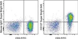
- Staining of normal human peripheral blood cells with Anti-Human CD3 FITC (Product # 11-0036-42) and Mouse IgG1 K Isotype Control Super Bright 645 (Product # 64-4714-82) (left) or Anti-Human CD127 Super Bright 645 (right). Cells in the lymphocyte gate were used for analysis.

Supportive validation
- Submitted by
- Invitrogen Antibodies (provider)
- Main image
- Experimental details
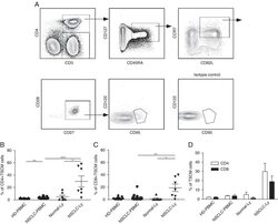
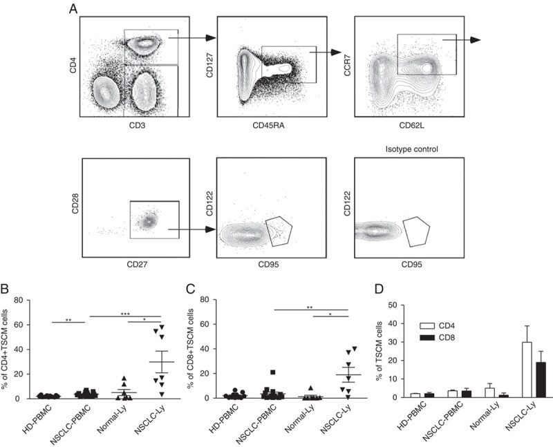
- FIGURE 1 Identification of Tscm CD4 + CD45RA + CD45RO - CD62L + CCR7 + CD127 + CD27 + CD28 + CD95 + CD122 + T (Tscm) cell in human blood and lymph nodes. PBMCs were isolated from the blood of non-small cell lung cancer (NSCLC) patients (n=15) (NSCLC-PBMC) and healthy donors (n=11) (HD-PBMC); lymphocytes were isolated from the tumor-infiltrated lymph node of NSCLC patients who were collected blood at same time (n=7) (NSCLC-Ly); lymphocytes were isolated from the healthy lymph node of non lung cancer patients (n=7) (Normal-Ly), analyzed by flow cytometry. A, Representative flow cytometric analyses of CD4 + CD45RA + CD45RO - CD62L + CCR7 + CD127 + CD27 + CD28 + CD95 + CD122 + T cells, indicating Tscm cells. B, The frequency of the CD4 + Tscm cells in the HD-PBMC, NSCLC-PBMC, Normal-Ly, NSCLC-Ly. The events of CD4 + Tscm cells in the blood and lymph node from NSCLC patients and healthy donors, expressed as the mean+-SEM. C, The frequency of the CD8 + Tscm cells in the HD-PBMC, NSCLC-PBMC, Normal-Ly, NSCLC-Ly, expressed as the mean+-SEM. D, The events of Tscm of CD4 + and CD8 + cells in the blood and the lymph node from NSCLC patients and healthy donors. HD indicates healthy donors; IFN, interferon; PBMC, peripheral blood mononuclear cells; Tscm cell, stem cell-like memory T cell. * P
- Submitted by
- Invitrogen Antibodies (provider)
- Main image
- Experimental details
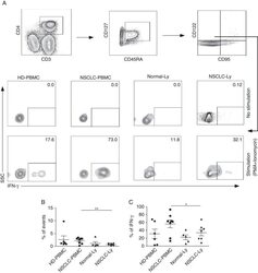
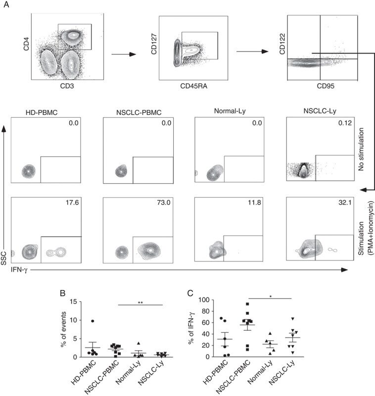
- FIGURE 3 CD4 + CD45RA + CD45RO - CD95 - CD122 + CD127 + T cell population displays different phenotypes in human blood and lymph nodes. A, Flow cytometry plots showing IFN-gamma expression in the CD4 + CD45RA + /CD45RO - CD95 - CD122 + CD127 + T cells from the blood and lymph node of the non-small cell lung cancer (NSCLC) patients and healthy donors. B, The mean frequency (+-SEM) of the events of CD4 + CD45RA + /CD45RO - CD95 - CD122 + CD127 + T cells. C, The mean IFN-gamma production (+-SEM) of the CD4 + CD45RA + /CD45RO - CD95 - CD122 + CD127 + T cells. HD indicates healthy donors; IFN, interferon; PBMC, peripheral blood mononuclear cells; PMA, phorbol 12-myristate13-acetate. * P
- Submitted by
- Invitrogen Antibodies (provider)
- Main image
- Experimental details
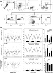
- Fig. 6 a Representative flow cytometry data, demonstrating the gating strategy used on PBMC for Treg identification and analysis. FSC-area vs. FSC-height was used for doublet discrimination. A ""dump"" channel was used to gate out dead cells (LIVE/DEAD fixable viability stain), CD14 + monocytes and CD19 + B cells, and lymphocytes were subsequently selected by FSC vs. SSC. CD4 + T cells were gated on the basis of CD4 vs. CD3 staining, then examined for expression of Ki67 and ICOS. Tregs were identified within the CD4 + T cell population as CD25 hi CD127 lo and Foxp3 + . b Longitudinal empirical data, linear mixed models and estimated means (left, centre and right-hand panels respectively) for Ki67+ and ICOS+ expression on CD4+ T cells, and the Treg proportion of CD4 cells ( P -values: *
