 Explore
 Validate
 Learn
Learn Immunocytochemistry
Antibody data
- Antibody Data
- Antigen structure
- References [1]
- Comments [0]
- Validations
- Immunocytochemistry [1]
- Immunohistochemistry [5]
- Other assay [1]
Submit
Validation data
Reference
Comment
Report error
- Product number
- PA5-64134 - Provider product page

- Provider
- Invitrogen Antibodies
- Product name
- MARCO Polyclonal Antibody
- Antibody type
- Polyclonal
- Antigen
- Recombinant protein fragment
- Description
- Immunogen sequence: LNLQARLRVL EMYFLNDTLA AEDSPSFSLL QSAHPGEHLA QGASRLQVLQ AQLTWVRV Highest antigen sequence identity to the following orthologs: Mouse - 52%, Rat - 57%.
- Reactivity
- Human
- Host
- Rabbit
- Isotype
- IgG
- Vial size
- 100 μL
- Concentration
- 0.30 mg/mL
- Storage
- Store at 4°C short term. For long term storage, store at -20°C, avoiding freeze/thaw cycles.
Submitted references Single-cell characterization of macrophages in glioblastoma reveals MARCO as a mesenchymal pro-tumor marker.
Chen AX, Gartrell RD, Zhao J, Upadhyayula PS, Zhao W, Yuan J, Minns HE, Dovas A, Bruce JN, Lasorella A, Iavarone A, Canoll P, Sims PA, Rabadan R
Genome medicine 2021 May 19;13(1):88
Genome medicine 2021 May 19;13(1):88
No comments: Submit comment
Supportive validation
- Submitted by
- Invitrogen Antibodies (provider)
- Main image
- Experimental details
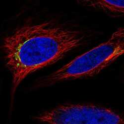
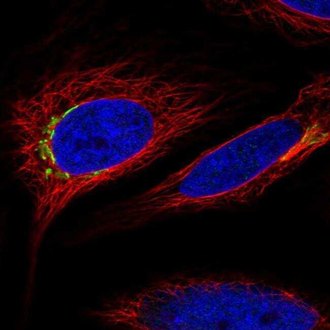
- Immunofluorescent staining of MARCO in human cell line HeLa using a MARCO Polyclonal Antibody (Product # PA5-64134) shows localization to the Golgi apparatus.
Supportive validation
- Submitted by
- Invitrogen Antibodies (provider)
- Main image
- Experimental details
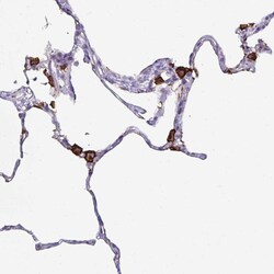
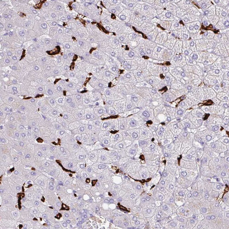
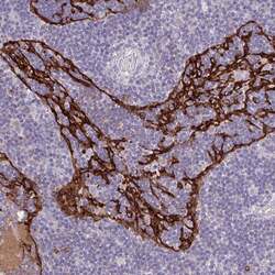
- Immunohistochemical staining of MARCO in human lung using MARCO Polyclonal Antibody (Product # PA5-64134) shows strong positivity in macrophages.

- Submitted by
- Invitrogen Antibodies (provider)
- Main image
- Experimental details
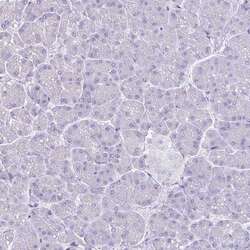
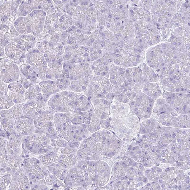
- Immunohistochemical staining of MARCO in human pancreas using MARCO Polyclonal Antibody (Product # PA5-64134) shows no positivity as expected.
- Submitted by
- Invitrogen Antibodies (provider)
- Main image
- Experimental details
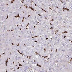
- Immunohistochemical staining of MARCO in human liver using MARCO Polyclonal Antibody (Product # PA5-64134) shows strong positivity in Kupffer cells.
- Submitted by
- Invitrogen Antibodies (provider)
- Main image
- Experimental details
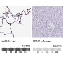
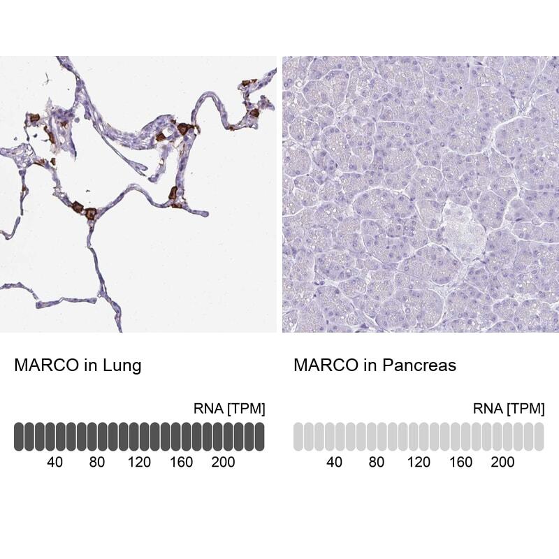
- Immunohistochemical staining of MARCO in human lung and pancreas tissues using MARCO Polyclonal Antibody (Product # PA5-64134). Corresponding MARCO RNA-seq data are presented for the same tissues.
- Submitted by
- Invitrogen Antibodies (provider)
- Main image
- Experimental details
- Immunohistochemical staining of MARCO in human lymph node using MARCO Polyclonal Antibody (Product # PA5-64134) shows moderate membranous positivity.

Supportive validation
- Submitted by
- Invitrogen Antibodies (provider)
- Main image
- Experimental details
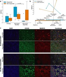
- Fig. 4 Dynamics of MARCO expression under PD1 immunotherapy. a Comparison of bulk MARCO expression in a longitudinal cohort of GBM patients treated with adjuvant PD1 checkpoint inhibitors following recurrence. Immunotherapy responders showed a decrease in MARCO expression between pre- and post-immunotherapy recurrences. b Timeline of representative examples of a responder (patient 55, blue) and non-responder (patient 20, orange) to PD1 immunotherapy. MARCO expression levels from available samples are plotted concurrently with the disease course. Each tick on the x -axis represents 1 month of time. c Immunofluorescence imaging of these same two representative cases (patients 55 and 20) before and after anti-PD-1 therapy, with dual staining of CD163 (green) and MARCO (red), alongside DAPI (blue)
