Explore
 Validate
 Learn
Learn Western blot
Antibody data
- Antibody Data
- Antigen structure
- References [0]
- Comments [0]
- Validations
- Western blot [1]
- Immunocytochemistry [2]
Submit
Validation data
Reference
Comment
Report error
- Product number
- PA5-77783 - Provider product page

- Provider
- Invitrogen Antibodies
- Product name
- RAB1B Polyclonal Antibody
- Antibody type
- Polyclonal
- Antigen
- Synthetic peptide
- Reactivity
- Human, Rat
- Host
- Rabbit
- Isotype
- IgG
- Vial size
- 100 µg
- Concentration
- 1 mg/mL
- Storage
- -20°C
No comments: Submit comment
Supportive validation
- Submitted by
- Invitrogen Antibodies (provider)
- Main image

- Experimental details
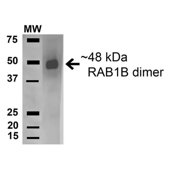
- Western blot analysis of RAB1B in human HeLa and 293T cell lysates with 15 µg of sample. The sample was blocked with 5% skim milk in TBST, incubated with RAB1B polyclonal antibody (Product # PA5-77783) using a dilution of 1:1000 (1 hour at RT), followed by Goat Anti-Rabbit HRP (1 hour at RT) at a dilution of 1:2000 and ECL development (6 min at RT). Samples were arranged as follows: Lane 1) Molecular Weight Ladder (MW), Lane 2) Human HeLa and 293Trap cell lysates.
Supportive validation
- Submitted by
- Invitrogen Antibodies (provider)
- Main image
- Experimental details
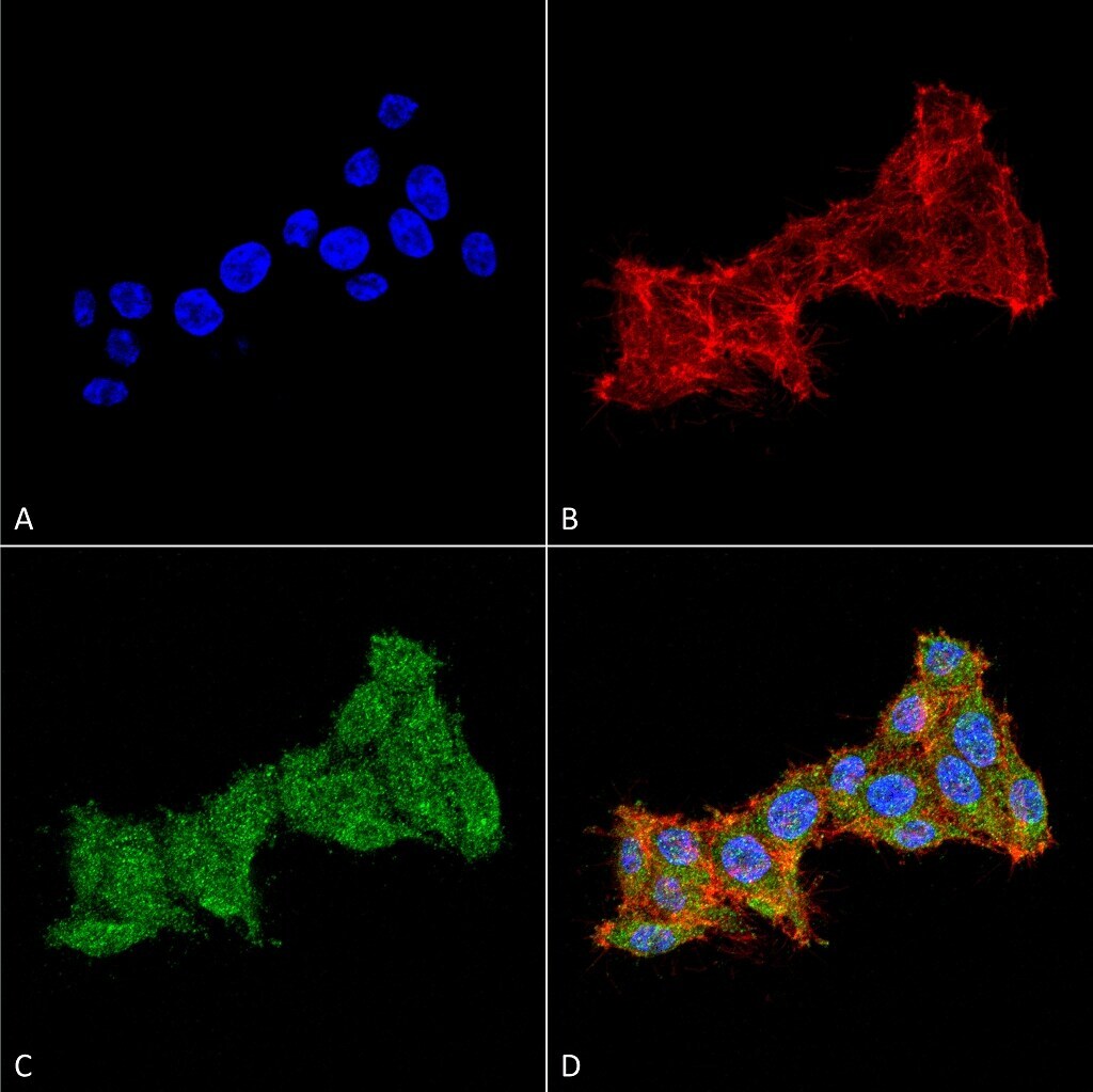
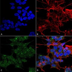
- Immunofluorescent analysis of RAB1B in human neuroblastoma cell line (SK-N-BE). Sample was fixed using 4% formaldehyde (15 min at RT), incubated with RAB1B polyclonal antibody (Product # PA5-77783) using a dilution of 1:100 (1 hour at RT), and followed by Goat Anti-Rabbit 488, Phalloidin Texas Red F-Actin stain, DAPI (blue) (1 hour at RT) at a dilution of 1:100, 1:1000 and 1:5000. Images shown are as follows: A) DAPI nuclear stain, B) Phalloidin Texas Red F-Actin stain, C) RAB1B Antibody, D) Composite. Magnification: 60X.
- Submitted by
- Invitrogen Antibodies (provider)
- Main image
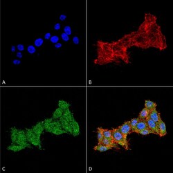
- Experimental details
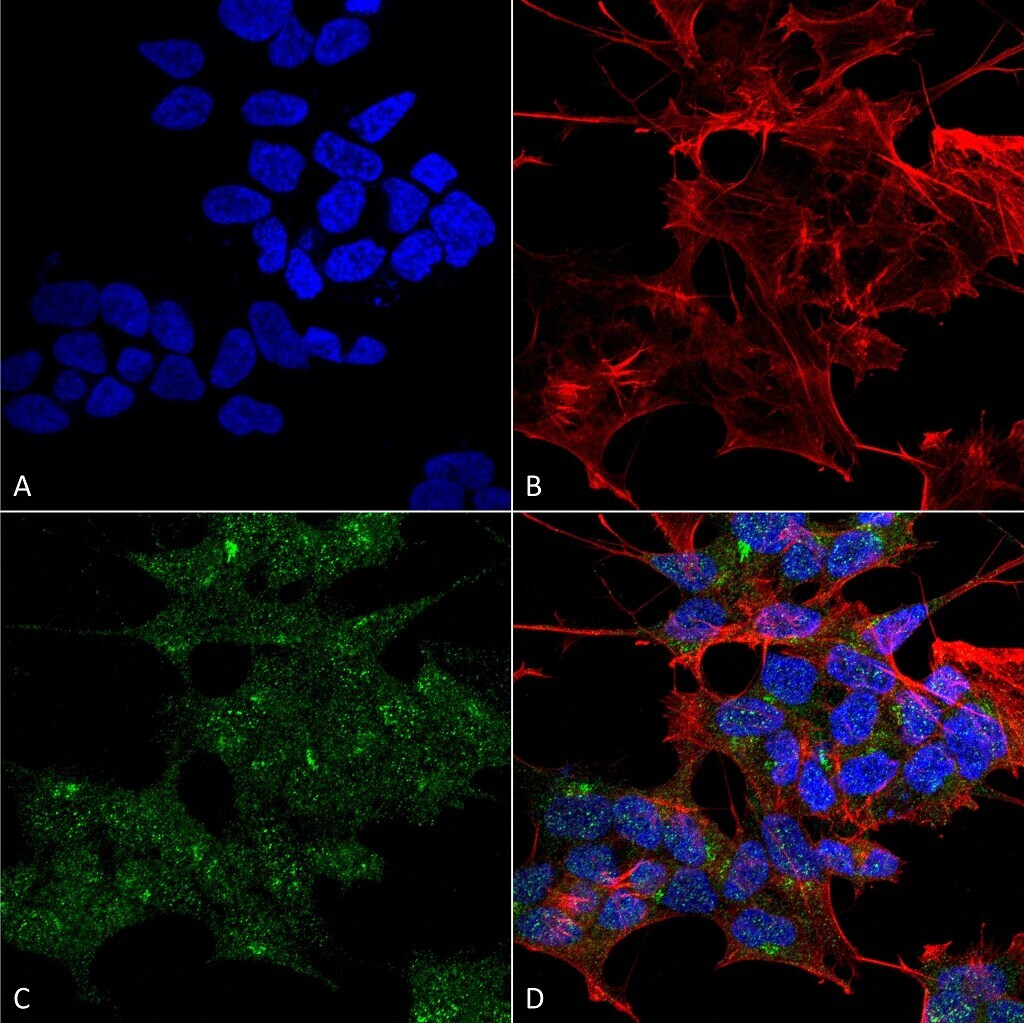
- Immunofluorescent analysis of RAB1B in human colon carcinoma cell line (RKO). Sample was fixed with 4% formaldehyde (15 min at RT), incubated with RAB1B polyclonal antibody (Product # PA5-77783) using a dilution of 1:100 (1 hour at RT), and followed by Goat Anti-Rabbit 488, Phalloidin Texas Red and DAPI secondary antibody at a dilution of 1:100, 1:1000 (60 min at RT) and 1:5000 (5 min at RT). Images are shown as follows: A) DAPI nuclear stain, B) Phalloidin Texas Red F-Actin stain, C) RAB1B Antibody, D) Merged image. Magnification: 60x.