Explore
 Validate
 Learn
Learn Western blot
Antibody data
- Antibody Data
- Antigen structure
- References [0]
- Comments [0]
- Validations
- Western blot [2]
- Immunocytochemistry [4]
- Immunohistochemistry [5]
- Flow cytometry [2]
- Chromatin Immunoprecipitation [2]
Submit
Validation data
Reference
Comment
Report error
- Product number
- MA5-32044 - Provider product page

- Provider
- Invitrogen Antibodies
- Product name
- PMS2 Recombinant Rabbit Monoclonal Antibody (SY08-09)
- Antibody type
- Monoclonal
- Antigen
- Synthetic peptide
- Description
- Recombinant rabbit monoclonal antibodies are produced using in vitro expression systems. The expression systems are developed by cloning in the specific antibody DNA sequences from immunoreactive rabbits. Then, individual clones are screened to select the best candidates for production. The advantages of using recombinant rabbit monoclonal antibodies include: better specificity and sensitivity, lot-to-lot consistency, animal origin-free formulations, and broader immunoreactivity to diverse targets due to larger rabbit immune repertoire.
- Reactivity
- Human
- Host
- Rabbit
- Isotype
- IgG
- Antibody clone number
- SY08-09
- Vial size
- 100 μL
- Concentration
- 1 mg/mL
- Storage
- Store at 4°C short term. For long term storage, store at -20°C, avoiding freeze/thaw cycles.
No comments: Submit comment
Supportive validation
- Submitted by
- Invitrogen Antibodies (provider)
- Main image
- Experimental details
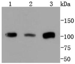
- Western blot analysis of PMS2 in different lysates using a Monoclonal antibody (Product #MA5-32044) at a dilution of 1:1,000. Positive control: Lane 1: Jurkat, Lane 2: Hela, Lane 3: SK-BR-3.

- Submitted by
- Invitrogen Antibodies (provider)
- Main image
- Experimental details
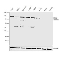
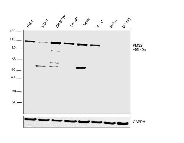
- Western blot was performed using Anti-PMS2 Recombinant Rabbit Monoclonal Antibody (SY08-09) (Product # MA5-32044) and a 95kDa band corresponding to PMS2 was observed in HeLa, MCF7, SH-SY5Y, LNCaP, Jurkat and PC-3 but not in Molt-4 and DU 145 which are reported to be negative. Whole cell extracts (30 µg lysate) of HeLa (Lane 1), MCF7 (Lane 2), SH-SY5Y (Lane 3), LnCaP (Lane 4), Jurkat (Lane 5), PC-3 (Lane 6), Molt-4 (Lane 7) and DU 145 (Lane 8) were electrophoresed using NuPAGE™ 4-12% Bis-Tris Protein Gel (Product # NP0322BOX). Resolved proteins were then transferred onto a nitrocellulose membrane (Product # IB23001) by iBlot® 2 Dry Blotting System (Product # IB21001). The blot was probed with the primary antibody (1:1000 dilution) and detected by chemiluminescence with Goat anti-Rabbit IgG (Heavy Chain), Superclonal™ Recombinant Secondary Antibody, HRP (Product # A27036, 1:4000 dilution) using the iBright FL 1000 (Product # A32752). Chemiluminescent detection was performed using Novex® ECL Chemiluminescent Substrate Reagent Kit (Product # WP20005).
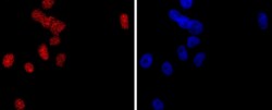
Supportive validation
- Submitted by
- Invitrogen Antibodies (provider)
- Main image

- Experimental details
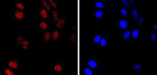
- Immunocytochemical analysis of PMS2 in Hela cells using a PMS2 Monoclonal antibody (Product # MA5-32044) as seen in red. The nuclear counter stain is DAPI (blue). Cells were fixed in paraformaldehyde, permeabilised with 0.25% Triton X100/PBS.
- Submitted by
- Invitrogen Antibodies (provider)
- Main image

- Experimental details
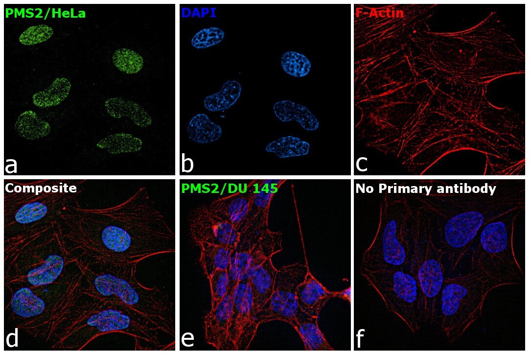
- Immunofluorescence analysis of PMS2 was performed using 70% confluent log phase HeLa and DU 145 cells. The cells were fixed with 4% paraformaldehyde for 10 minutes, permeabilized with 0.1% Triton™ X-100 for 15 minutes, and blocked with 2% BSA for 1 hour at room temperature. HeLa cells were labeled with PMS2 Rabbit Monoclonal Antibody (Product # MA5-32044) at 1:250 dilution in 0.1% BSA, incubated at 4 degree Celsius overnight and then labeled with Goat anti-Rabbit IgG (H+L) Superclonal™ Recombinant Secondary Antibody, Alexa Fluor® 488 conjugate (Product # A27034) at a dilution of 1:2000 for 45 minutes at room temperature (Panel a: green). Nuclei (Panel b: blue) were stained with ProLong™ Diamond Antifade Mountant with DAPI (Product # P36962). F-actin (Panel c: red) was stained with Rhodamine Phalloidin (Product # R415). Panel d represents the merged image of HeLa showing nuclear localization. Panel e represents the merged image of DU 145 cells showing no expression for PMS2 protein. Panel f represents control cells with no primary antibody to assess background. The images were captured at 60X magnification.
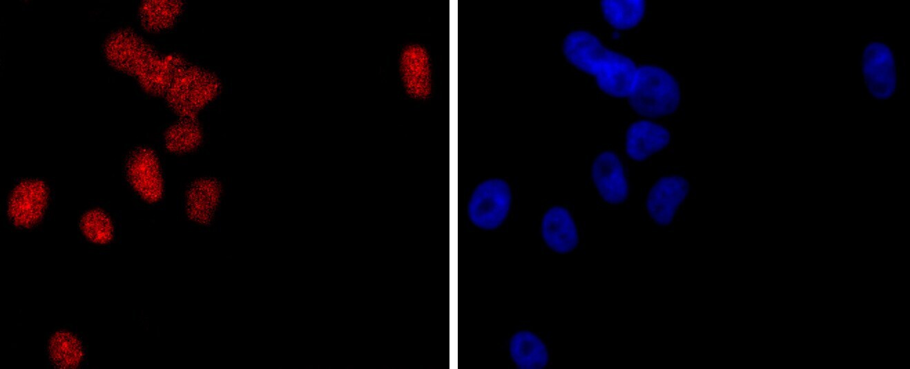
- Submitted by
- Invitrogen Antibodies (provider)
- Main image
- Experimental details
- Immunocytochemical analysis of PMS2 in Hela cells using a PMS2 Monoclonal antibody (Product # MA5-32044) as seen in red. The nuclear counter stain is DAPI (blue). Cells were fixed in paraformaldehyde, permeabilised with 0.25% Triton X100/PBS.

- Submitted by
- Invitrogen Antibodies (provider)
- Main image
- Experimental details
- Immunofluorescence analysis of PMS2 was performed using 70% confluent log phase HeLa and DU 145 cells. The cells were fixed with 4% paraformaldehyde for 10 minutes, permeabilized with 0.1% Triton™ X-100 for 15 minutes, and blocked with 2% BSA for 1 hour at room temperature. HeLa cells were labeled with PMS2 Rabbit Monoclonal Antibody (Product # MA5-32044) at 1:250 dilution in 0.1% BSA, incubated at 4 degree Celsius overnight and then labeled with Goat anti-Rabbit IgG (Heavy Chain) Superclonal™ Recombinant Secondary Antibody, Alexa Fluor® 488 conjugate (Product # A27034) at a dilution of 1:2000 for 45 minutes at room temperature (Panel a: green). Nuclei (Panel b: blue) were stained with ProLong™ Diamond Antifade Mountant with DAPI (Product # P36962). F-actin (Panel c: red) was stained with Rhodamine Phalloidin (Product # R415). Panel d represents the merged image of HeLa showing nuclear localization. Panel e represents the merged image of DU 145 cells showing no expression for PMS2 protein. Panel f represents control cells with no primary antibody to assess background. The images were captured at 60X magnification.

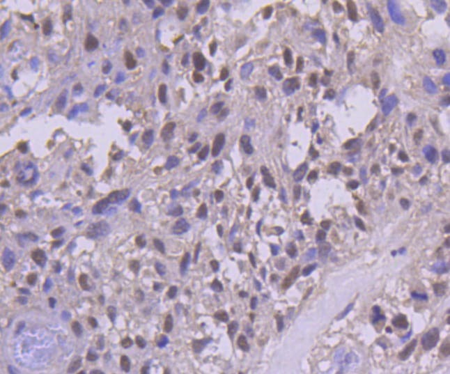
Supportive validation
- Submitted by
- Invitrogen Antibodies (provider)
- Main image
- Experimental details
- Immunohistochemical analysis of PMS2 of paraffin-embedded Human breast carcinoma tissue using a PMS2 Monoclonal antibody (Product #MA5-32044). Counter stained with hematoxylin.
- Submitted by
- Invitrogen Antibodies (provider)
- Main image

- Experimental details
- Immunohistochemical analysis of PMS2 of paraffin-embedded Human breast carcinoma tissue using a PMS2 Monoclonal antibody (Product #MA5-32044). Counter stained with hematoxylin..

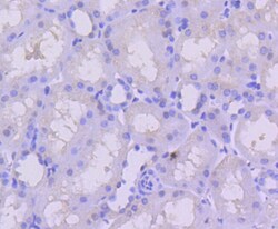
- Submitted by
- Invitrogen Antibodies (provider)
- Main image

- Experimental details
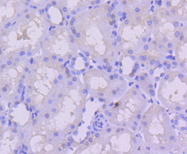
- Immunohistochemical analysis of PMS2 of paraffin-embedded Human kidney tissue using a PMS2 Monoclonal antibody (Product #MA5-32044). Counter stained with hematoxylin..

- Submitted by
- Invitrogen Antibodies (provider)
- Main image

- Experimental details
- Immunohistochemical analysis of PMS2 of paraffin-embedded Human breast carcinoma tissue using a PMS2 Monoclonal antibody (Product #MA5-32044). Counter stained with hematoxylin..
- Submitted by
- Invitrogen Antibodies (provider)
- Main image
- Experimental details
- Immunohistochemical analysis of PMS2 of paraffin-embedded Human kidney tissue using a PMS2 Monoclonal antibody (Product #MA5-32044). Counter stained with hematoxylin..
Supportive validation
- Submitted by
- Invitrogen Antibodies (provider)
- Main image
- Experimental details
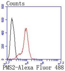
- Flow Cytometric analysis of PMS2 in Hela cells using a PMS2 Monoclonal Antibody (Product # MA5-32044) at a dilution of 1:50, as seen in red compared with an unlabelled control (cells without incubation with primary antibody; black). Alexa Fluor 488-conjugated goat anti rabbit IgG was used as the secondary antibody..

- Submitted by
- Invitrogen Antibodies (provider)
- Main image

- Experimental details
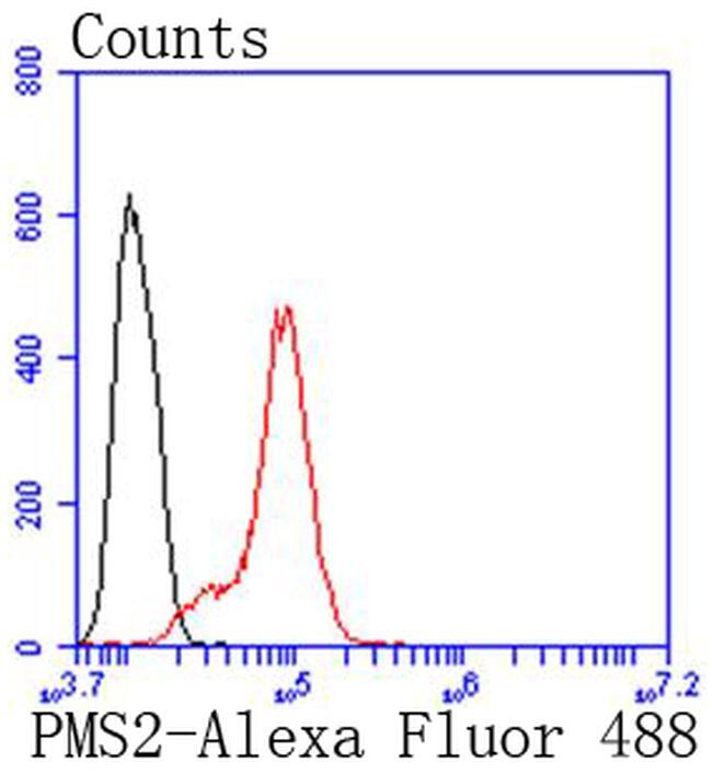
- Flow Cytometric analysis of PMS2 in Hela cells using a PMS2 Monoclonal Antibody (Product # MA5-32044) at a dilution of 1:50, as seen in red compared with an unlabelled control (cells without incubation with primary antibody; black). Alexa Fluor 488-conjugated goat anti rabbit IgG was used as the secondary antibody..
Supportive validation
- Submitted by
- Invitrogen Antibodies (provider)
- Main image

- Experimental details
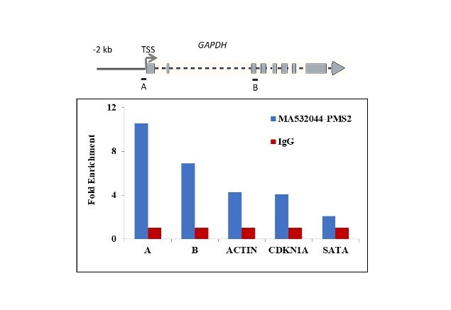
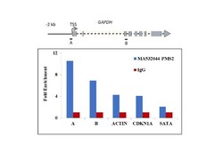
- Chromatin Immunoprecipitation (ChIP) assay of endogenous PMS2 protein using Anti-PMS2 Antibody: ChIP was performed using Anti-PMS2 Rabbit Recombinant Monoclonal Antibody (Product # MA5-32044, 5 µg) on sheared chromatin from methyl methanesulfonate treated SW480 cells using the MAGnify ChIP System kit (Product # 49-2024). Normal Rabbit IgG was used as a negative IP control. The purified DNA was analyzed by qPCR using primers binding to GAPDH transcriptional start site, GAPDH gene body (+2Kb), ACTB promoter, CDKN1A intron 1 and SATA satellite repeats. Data is presented as fold enrichment of the antibody signal versus the negative control IgG using the comparative CT method.

- Submitted by
- Invitrogen Antibodies (provider)
- Main image
- Experimental details
- Chromatin Immunoprecipitation (ChIP) assay of endogenous PMS2 protein using Anti-PMS2 Antibody: ChIP was performed using Anti-PMS2 Rabbit Recombinant Monoclonal Antibody (Product # MA5-32044, 5 µg) on sheared chromatin from methyl methanesulfonate treated SW480 cells using the MAGnify ChIP System kit (Product # 49-2024). Normal Rabbit IgG was used as a negative IP control. The purified DNA was analyzed by qPCR using primers binding to GAPDH transcriptional start site, GAPDH gene body (+2Kb), ACTB promoter, CDKN1A intron 1 and SATA satellite repeats. Data is presented as fold enrichment of the antibody signal versus the negative control IgG using the comparative CT method.