Explore
 Validate
 Learn
Learn Flow cytometry
Antibody data
- Antibody Data
- Antigen structure
- References [4]
- Comments [0]
- Validations
- Flow cytometry [1]
- Other assay [1]
Submit
Validation data
Reference
Comment
Report error
- Product number
- 13-1319-82 - Provider product page

- Provider
- Invitrogen Antibodies
- Product name
- CD131 Monoclonal Antibody (1C1), Biotin, eBioscience™
- Antibody type
- Monoclonal
- Antigen
- Other
- Description
- Description: The 1C1 monoclonal antibody reacts with the human CD131 molecule, also known as the common beta subunit (betaC). The common beta subunit associates with the specific alpha subunits of IL-3 receptor, IL-5 receptor and GM-CSF receptor to form high affinity receptors for these cytokines. These cytokine receptors are expressed by neutrophils, eosinophils, monocytes, endothelial cells, fibroblasts and hematopoietic progenitor cells and play a crucial role in growth/activation of eosinophils and in the inflammatory response. Applications Reported: This 1C1 antibody has been reported for use in flow cytometric analysis. Applications Tested: This 1C1 antibody has been tested by flow cytometric analysis of normal human peripheral blood cells. This can be used at less than or equal to 0.5 µg per test. A test is defined as the amount (µg) of antibody that will stain a cell sample in a final volume of 100 µL. Cell number should be determined empirically but can range from 10^5 to 10^8 cells/test. It is recommended that the antibody be carefully titrated for optimal performance in the assay of interest. Filtration: 0.2 µm post-manufacturing filtered.
- Reactivity
- Human
- Host
- Mouse
- Conjugate
- Biotin
- Isotype
- IgG
- Antibody clone number
- 1C1
- Vial size
- 100 μg
- Concentration
- 0.5 mg/mL
- Storage
- 4°C, store in dark, DO NOT FREEZE!
Submitted references Severe T cell hyporeactivity in ventilated COVID-19 patients correlates with prolonged virus persistence and poor outcomes.
Simultaneous antagonism of interleukin-5, granulocyte-macrophage colony-stimulating factor, and interleukin-3 stimulation of human eosinophils by targetting the common cytokine binding site of their receptors.
The human granulocyte-macrophage colony-stimulating factor (GM-CSF) receptor exists as a preformed receptor complex that can be activated by GM-CSF, interleukin-3, or interleukin-5.
Interleukin-5, interleukin-3, and granulocyte-macrophage colony-stimulating factor cross-compete for binding to cell surface receptors on human eosinophils.
Renner K, Schwittay T, Chaabane S, Gottschling J, Müller C, Tiefenböck C, Salewski JN, Winter F, Buchtler S, Balam S, Malfertheiner MV, Lubnow M, Lunz D, Graf B, Hitzenbichler F, Hanses F, Poeck H, Kreutz M, Orsó E, Burkhardt R, Niedermair T, Brochhausen C, Gessner A, Salzberger B, Mack M
Nature communications 2021 May 21;12(1):3006
Nature communications 2021 May 21;12(1):3006
Simultaneous antagonism of interleukin-5, granulocyte-macrophage colony-stimulating factor, and interleukin-3 stimulation of human eosinophils by targetting the common cytokine binding site of their receptors.
Sun Q, Jones K, McClure B, Cambareri B, Zacharakis B, Iversen PO, Stomski F, Woodcock JM, Bagley CJ, D'Andrea R, Lopez AF
Blood 1999 Sep 15;94(6):1943-51
Blood 1999 Sep 15;94(6):1943-51
The human granulocyte-macrophage colony-stimulating factor (GM-CSF) receptor exists as a preformed receptor complex that can be activated by GM-CSF, interleukin-3, or interleukin-5.
Woodcock JM, McClure BJ, Stomski FC, Elliott MJ, Bagley CJ, Lopez AF
Blood 1997 Oct 15;90(8):3005-17
Blood 1997 Oct 15;90(8):3005-17
Interleukin-5, interleukin-3, and granulocyte-macrophage colony-stimulating factor cross-compete for binding to cell surface receptors on human eosinophils.
Lopez AF, Vadas MA, Woodcock JM, Milton SE, Lewis A, Elliott MJ, Gillis D, Ireland R, Olwell E, Park LS
The Journal of biological chemistry 1991 Dec 25;266(36):24741-7
The Journal of biological chemistry 1991 Dec 25;266(36):24741-7
No comments: Submit comment
Supportive validation
- Submitted by
- Invitrogen Antibodies (provider)
- Main image
- Experimental details
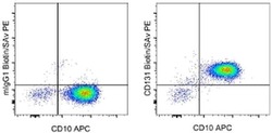
- Staining of normal human peripheral blood cells with Anti-Human CD10 APC (Product # 17-0106) and 0.25 µg of Mouse IgG1 K Isotype Control Biotin (Product # 13-4714) (left) or 0.25 µg of Anti-Human CD131 Biotin (right) followed by Streptavidin PE (Product # 12-4317). Cells in the granulocyte gate were used for analysis.

Supportive validation
- Submitted by
- Invitrogen Antibodies (provider)
- Main image
- Experimental details
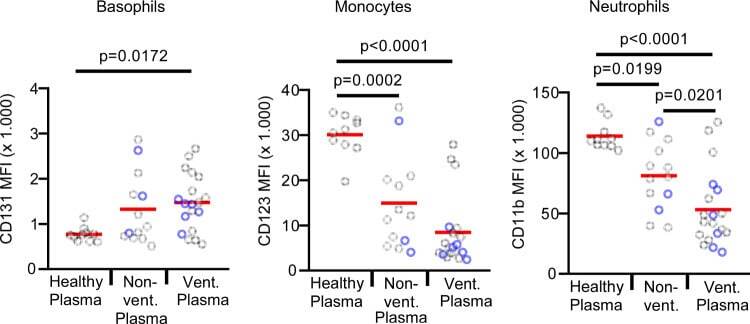
- Fig. 3 Plasma from COVID-19 patients has pronounced T-cell suppressive activity. Whole blood from a healthy donor was washed twice with medium to remove the plasma. Plasma from 10 healthy controls (healthy plasma; n = 10 biologically independent samples), 13 non-ventilated (non-vent. plasma; n = 13 biologically independent samples), and 15 mechanically ventilated (vent. plasma; n = 19 biologically independent samples) COVID-19 patients was added and samples were cultured with anti-CD3 for 24 h. In all, 23% of the non-ventilated and 33% of the ventilated COVID-19 patients were treated with steroids (marked in blue). None of the patients was treated with other immunosuppressive agents. Expression of indicated surface markers was quantified by flow cytometry on basophils, CD14 + monocytes and neutrophils. The absolute expression values of indicated markers are shown as mean fluorescence intensity (MFI) on basophils, CD14 + monocytes, and neutrophils. Each sample is represented by one dot, and the mean is marked in red. One-way ANOVA with Bonferroni multiple comparison test was used. Source data are provided as a Source Data file.
- Conjugate
- Biotin