Explore
 Validate
 Learn
Learn Western blot
 Immunocytochemistry
Antibody data
- Antibody Data
- Antigen structure
- References [1]
- Comments [0]
- Validations
- Immunocytochemistry [3]
- Immunohistochemistry [1]
- Other assay [1]
Submit
Validation data
Reference
Comment
Report error
- Product number
- PA5-37601 - Provider product page

- Provider
- Invitrogen Antibodies
- Product name
- Phospho-IGF1R beta (Tyr1161) Polyclonal Antibody
- Antibody type
- Polyclonal
- Antigen
- Synthetic peptide
- Description
- A suggested positive control for Western blot is 293 cells; suggested positive control for IHC is human breast carcinoma; suggested positive control for ICC/IF is MCF-7 cells.
- Reactivity
- Human, Mouse, Rat
- Host
- Rabbit
- Isotype
- IgG
- Vial size
- 100 μL
- Concentration
- 1 mg/mL
- Storage
- -20°C
Submitted references Disc1 Carrier Mice Exhibit Alterations in Neural pIGF-1Rβ and Related Kinase Expression.
Sultana R, Shrestha A, Lee CC, Ogundele OM
Frontiers in cellular neuroscience 2020;14:94
Frontiers in cellular neuroscience 2020;14:94
No comments: Submit comment
Supportive validation
- Submitted by
- Invitrogen Antibodies (provider)
- Main image
- Experimental details
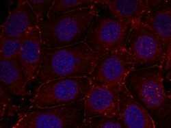
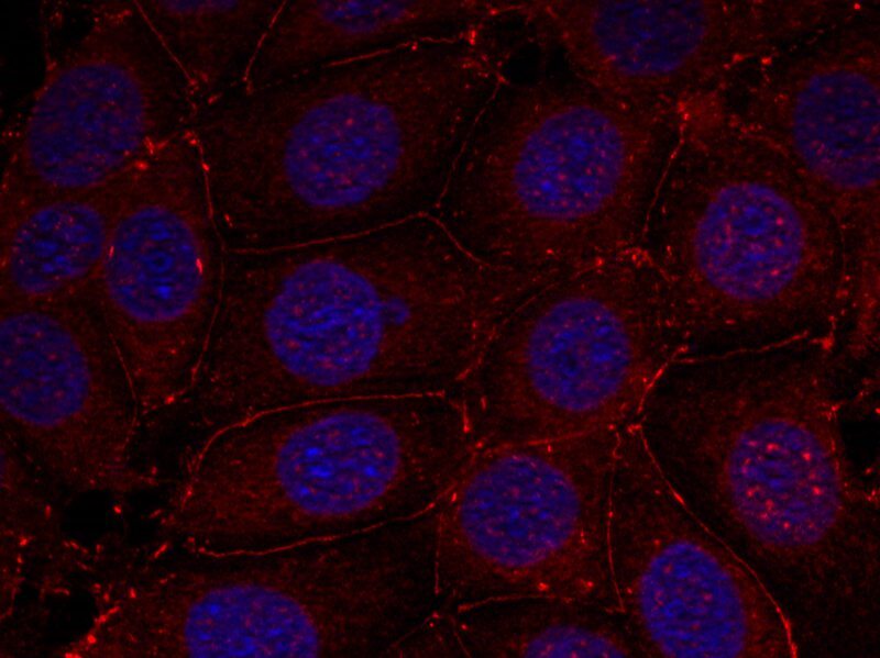
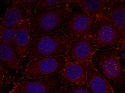
- Immunofluorescence staining of methanol-fixed MCF-7 cells using IGF-1R (pTyr1161) polyclonal antibody (Product # PA5-37601) (red).

- Submitted by
- Invitrogen Antibodies (provider)
- Main image

- Experimental details
- Immunocytochemical analysis of Phospho-IGF1R beta (Tyr1161) in methanol-fixed MCF7 cells, using Phospho-IGF1R beta (Tyr1161) Polyclonal Antibody (Product # PA5-37601)(Red).
- Submitted by
- Invitrogen Antibodies (provider)
- Main image
- Experimental details
- Immunocytochemical analysis of Phospho-IGF1R beta (Tyr1161) in methanol-fixed MCF7 cells, using Phospho-IGF1R beta (Tyr1161) Polyclonal Antibody (Product # PA5-37601)(Red).

Supportive validation
- Submitted by
- Invitrogen Antibodies (provider)
- Main image
- Experimental details
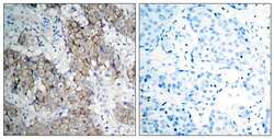
- Immunohistochemical analysis of Phospho-IGF1R beta (Tyr1161) in paraffin-embedded Human breast carcinoma tissue using Phospho-IGF1R beta (Tyr1161) Polyclonal Antibody (Product # PA5-37601).

Supportive validation
- Submitted by
- Invitrogen Antibodies (provider)
- Main image
- Experimental details
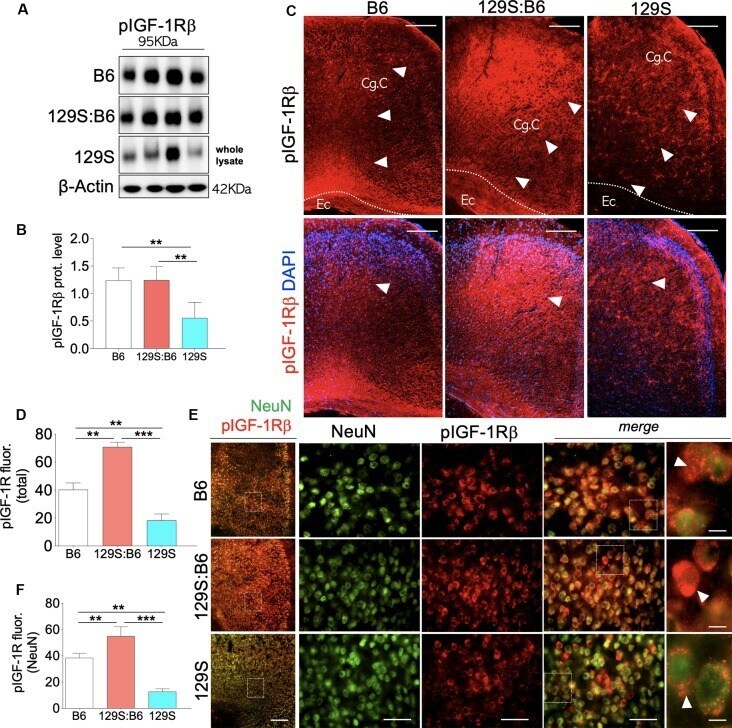
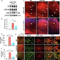
- Figure 3 Prefrontal cortex (PFC) expression of pIGF-1Rbeta in 129S:B6 and 129S mice. (A) Representative western blots for pIGF-1Rbeta detection in prefrontal cortical whole tissue lysate. (B) One-Way ANOVA comparison of normalized cortical pIGF-1Rbeta expression. (C) Low magnification immunofluorescence images demonstrating an increase in pIGF-1Rbeta expression in the 129S:B6 PFC, and a decrease in the 129S cortex (scale bar = 200 mum; Cg. C: cingulate cortex and Ec: external capsule). (D) Bar graph comparing pIGF-1Rbeta fluorescence intensity in the PFC. (E) Double fluorescence immunolabeling for NeuN/pIGF-1Rbeta co-localization in the PFC (scale bar = 200 mum, 60 mum, and 10 mum). (F) Quantification of pIGF-1Rbeta fluorescence intensity in NeuN-labeled PFC neurons [( n = 4 to n = 6 per group; B,D,F ); ** p < 0.01 and *** p < 0.001].