Explore
 Validate
 Learn
Learn44-684G
antibody from Invitrogen Antibodies
Targeting: MAPK14
CSBP1, CSBP2, CSPB1, Mxi2, p38, PRKM14, PRKM15
 Western blot
 Immunocytochemistry
Antibody data
- Antibody Data
- Antigen structure
- References [28]
- Comments [0]
- Validations
- Immunocytochemistry [4]
- Immunohistochemistry [3]
- Flow cytometry [1]
- Other assay [6]
Submit
Validation data
Reference
Comment
Report error
- Product number
- 44-684G - Provider product page

- Provider
- Invitrogen Antibodies
- Product name
- Phospho-p38 MAPK (Thr180, Tyr182) Polyclonal Antibody
- Antibody type
- Polyclonal
- Antigen
- Synthetic peptide
- Description
- This antibody reacts with human and rat (100% homologous) p38 alpha, beta, and gamma. HEK 293 cells +/- UV irradiation treatment and PC12 cells +/- Sorbitol were used as positive controls. Purified from rabbit serum by sequential epitope-specific chromatography, this product contains enough material for 10 mini-blots. The antibody has been negatively preadsorbed using a non-phosphopeptide corresponding to the site of phosphorylation to remove antibody that is reactive with non-phosphorylated p38. The final product is generated by affinity chromatography using a p38-derived peptide that is phosphorylated at threonine 180 and tyrosine 182.
- Reactivity
- Human, Rat
- Host
- Rabbit
- Isotype
- IgG
- Vial size
- 100 μL
- Storage
- -20°C
Submitted references Downregulation of FPR1 abates lipopolysaccharide-induced inflammatory injury and apoptosis by upregulating MAPK signaling pathway in murine chondrogenic ATDC5 cells.
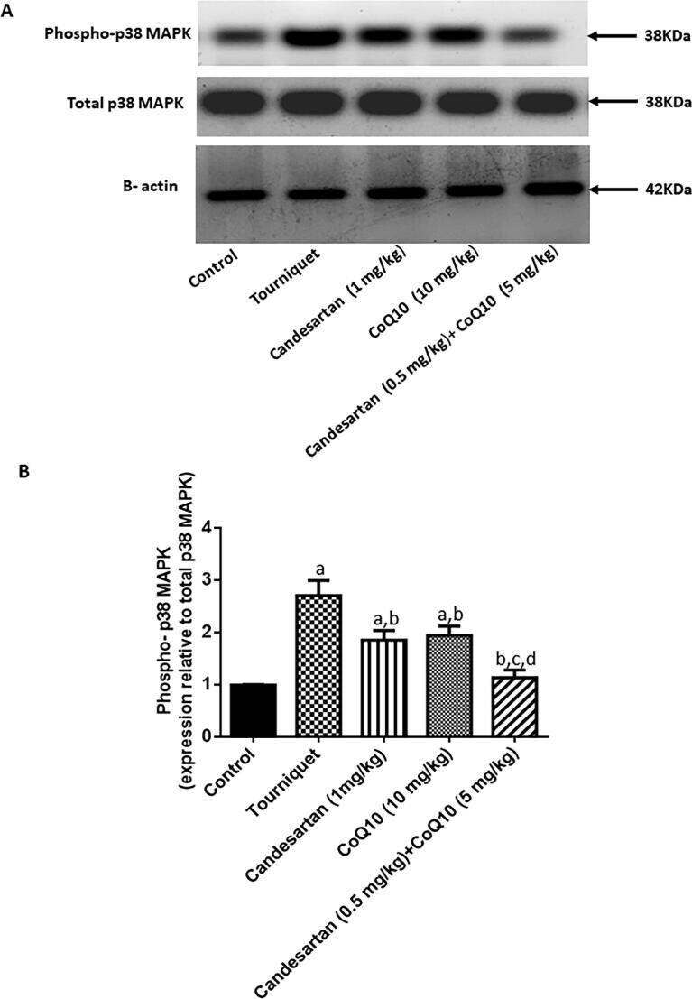
CoQ10 augments candesartan protective effect against tourniquet-induced hind limb ischemia-reperfusion: Involvement of non-classical RAS and ROS pathways.
Evidence for acupoint catgut embedding treatment and TRPV1 gene deletion increasing weight control in murine model.
A novel protective role of sacubitril/valsartan in cyclophosphamide induced lung injury in rats: impact of miRNA-150-3p on NF-κB/MAPK signaling trajectories.
Whole Genome Microarray Analysis of DUSP4-Deletion Reveals A Novel Role for MAP Kinase Phosphatase-2 (MKP-2) in Macrophage Gene Expression and Function.
Hypo-phosphorylated CD147 promotes migration and invasion of hepatocellular carcinoma cells and predicts a poor prognosis.
RARβ2-dependent signaling represses neuronal differentiation in mouse ES cells.
Exosomal miR-141-3p regulates osteoblast activity to promote the osteoblastic metastasis of prostate cancer.
12-Lipoxygenase Inhibitor Improves Functions of Cytokine-Treated Human Islets and Type 2 Diabetic Islets.
Cathepsin S attenuates endosomal EGFR signalling: A mechanical rationale for the combination of cathepsin S and EGFR tyrosine kinase inhibitors.
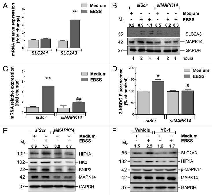
MAPK14/p38α-dependent modulation of glucose metabolism affects ROS levels and autophagy during starvation.
The genomic landscape of diffuse intrinsic pontine glioma and pediatric non-brainstem high-grade glioma.
Expansion of oligodendrocyte progenitor cells following SIRT1 inactivation in the adult brain.
Molecular pathway profiling of T lymphocyte signal transduction pathways; Th1 and Th2 genomic fingerprints are defined by TCR and CD28-mediated signaling.
Growth-factor receptor-bound protein-2 (Grb2) signaling in B cells controls lymphoid follicle organization and germinal center reaction.
T-cell receptor ligation induces distinct signaling pathways in naive vs. antigen-experienced T cells.
Differential modulation of TLR3- and TLR4-mediated dendritic cell maturation and function by progesterone.
TGFbeta1 antagonistic peptides inhibit TGFbeta1-dependent angiogenesis.
Chemotherapeutic agents up-regulate the cytomegalovirus promoter: implications for bioluminescence imaging of tumor response to therapy.
Molecular mechanisms involved in interleukin-4-induced human neutrophils: expression and regulation of suppressor of cytokine signaling.
Critical role of c-jun (NH2) terminal kinase in paracetamol- induced acute liver failure.
The peroxisome proliferator-activated receptor gamma agonist pioglitazone reduces the development of cartilage lesions in an experimental dog model of osteoarthritis: in vivo protective effects mediated through the inhibition of key signaling and catabolic pathways.
The mitogen-activated protein kinases (MAPK) p38 and JNK are markers of tumor progression in breast carcinoma.
TNF-alpha promotes a stop signal that inhibits neutrophil polarization and migration via a p38 MAPK pathway.
Plasmin induces smooth muscle cell proliferation.
Protein synthesis persists during necrotic cell death.
Chemokine receptor CCR2 expression by systemic sclerosis fibroblasts: evidence for autocrine regulation of myofibroblast differentiation.
Urokinase-induced smooth muscle cell responses require distinct signaling pathways: a role for the epidermal growth factor receptor.
Chen H, Zhang L
Allergologia et immunopathologia 2021;49(5):57-63
Allergologia et immunopathologia 2021;49(5):57-63
CoQ10 augments candesartan protective effect against tourniquet-induced hind limb ischemia-reperfusion: Involvement of non-classical RAS and ROS pathways.
Awad AS, Nour El-Din M, Kamel R
Saudi pharmaceutical journal : SPJ : the official publication of the Saudi Pharmaceutical Society 2021 Jul;29(7):724-733
Saudi pharmaceutical journal : SPJ : the official publication of the Saudi Pharmaceutical Society 2021 Jul;29(7):724-733
Evidence for acupoint catgut embedding treatment and TRPV1 gene deletion increasing weight control in murine model.
Inprasit C, Huang YC, Lin YW
International journal of molecular medicine 2020 Mar;45(3):779-792
International journal of molecular medicine 2020 Mar;45(3):779-792
A novel protective role of sacubitril/valsartan in cyclophosphamide induced lung injury in rats: impact of miRNA-150-3p on NF-κB/MAPK signaling trajectories.
Abdel-Latif GA, Elwahab AHA, Hasan RA, ElMongy NF, Ramzy MM, Louka ML, Schaalan MF
Scientific reports 2020 Aug 3;10(1):13045
Scientific reports 2020 Aug 3;10(1):13045
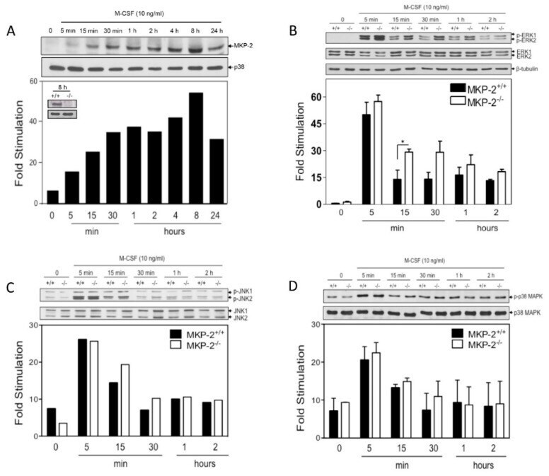
Whole Genome Microarray Analysis of DUSP4-Deletion Reveals A Novel Role for MAP Kinase Phosphatase-2 (MKP-2) in Macrophage Gene Expression and Function.
Neamatallah T, Jabbar S, Tate R, Schroeder J, Shweash M, Alexander J, Plevin R
International journal of molecular sciences 2019 Jul 12;20(14)
International journal of molecular sciences 2019 Jul 12;20(14)
Hypo-phosphorylated CD147 promotes migration and invasion of hepatocellular carcinoma cells and predicts a poor prognosis.
Jin J, Wang SJ, Cui J, Li L, Li JY, Liu FL, Sun XX, Jiang JL, Cui HY, Chen ZN
Cellular oncology (Dordrecht) 2019 Aug;42(4):537-554
Cellular oncology (Dordrecht) 2019 Aug;42(4):537-554
RARβ2-dependent signaling represses neuronal differentiation in mouse ES cells.
Kona SL, Shrestha A, Yi X, Joseph S, Barona HM, Martinez-Ceballos E
Differentiation; research in biological diversity 2017 Nov-Dec;98:55-61
Differentiation; research in biological diversity 2017 Nov-Dec;98:55-61
Exosomal miR-141-3p regulates osteoblast activity to promote the osteoblastic metastasis of prostate cancer.
Ye Y, Li SL, Ma YY, Diao YJ, Yang L, Su MQ, Li Z, Ji Y, Wang J, Lei L, Fan WX, Li LX, Xu Y, Hao XK
Oncotarget 2017 Nov 7;8(55):94834-94849
Oncotarget 2017 Nov 7;8(55):94834-94849
12-Lipoxygenase Inhibitor Improves Functions of Cytokine-Treated Human Islets and Type 2 Diabetic Islets.
Ma K, Xiao A, Park SH, Glenn L, Jackson L, Barot T, Weaver JR, Taylor-Fishwick DA, Luci DK, Maloney DJ, Mirmira RG, Imai Y, Nadler JL
The Journal of clinical endocrinology and metabolism 2017 Aug 1;102(8):2789-2797
The Journal of clinical endocrinology and metabolism 2017 Aug 1;102(8):2789-2797
Cathepsin S attenuates endosomal EGFR signalling: A mechanical rationale for the combination of cathepsin S and EGFR tyrosine kinase inhibitors.
Huang CC, Lee CC, Lin HH, Chang JY
Scientific reports 2016 Jul 8;6:29256
Scientific reports 2016 Jul 8;6:29256
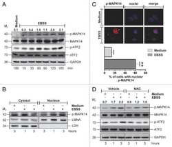
MAPK14/p38α-dependent modulation of glucose metabolism affects ROS levels and autophagy during starvation.
Desideri E, Vegliante R, Cardaci S, Nepravishta R, Paci M, Ciriolo MR
Autophagy 2014 Sep;10(9):1652-65
Autophagy 2014 Sep;10(9):1652-65
The genomic landscape of diffuse intrinsic pontine glioma and pediatric non-brainstem high-grade glioma.
Wu G, Diaz AK, Paugh BS, Rankin SL, Ju B, Li Y, Zhu X, Qu C, Chen X, Zhang J, Easton J, Edmonson M, Ma X, Lu C, Nagahawatte P, Hedlund E, Rusch M, Pounds S, Lin T, Onar-Thomas A, Huether R, Kriwacki R, Parker M, Gupta P, Becksfort J, Wei L, Mulder HL, Boggs K, Vadodaria B, Yergeau D, Russell JC, Ochoa K, Fulton RS, Fulton LL, Jones C, Boop FA, Broniscer A, Wetmore C, Gajjar A, Ding L, Mardis ER, Wilson RK, Taylor MR, Downing JR, Ellison DW, Zhang J, Baker SJ
Nature genetics 2014 May;46(5):444-450
Nature genetics 2014 May;46(5):444-450
Expansion of oligodendrocyte progenitor cells following SIRT1 inactivation in the adult brain.
Rafalski VA, Ho PP, Brett JO, Ucar D, Dugas JC, Pollina EA, Chow LM, Ibrahim A, Baker SJ, Barres BA, Steinman L, Brunet A
Nature cell biology 2013 Jun;15(6):614-24
Nature cell biology 2013 Jun;15(6):614-24
Molecular pathway profiling of T lymphocyte signal transduction pathways; Th1 and Th2 genomic fingerprints are defined by TCR and CD28-mediated signaling.
Smeets RL, Fleuren WW, He X, Vink PM, Wijnands F, Gorecka M, Klop H, Bauerschmidt S, Garritsen A, Koenen HJ, Joosten I, Boots AM, Alkema W
BMC immunology 2012 Mar 14;13:12
BMC immunology 2012 Mar 14;13:12
Growth-factor receptor-bound protein-2 (Grb2) signaling in B cells controls lymphoid follicle organization and germinal center reaction.
Jang IK, Cronshaw DG, Xie LK, Fang G, Zhang J, Oh H, Fu YX, Gu H, Zou Y
Proceedings of the National Academy of Sciences of the United States of America 2011 May 10;108(19):7926-31
Proceedings of the National Academy of Sciences of the United States of America 2011 May 10;108(19):7926-31
T-cell receptor ligation induces distinct signaling pathways in naive vs. antigen-experienced T cells.
Adachi K, Davis MM
Proceedings of the National Academy of Sciences of the United States of America 2011 Jan 25;108(4):1549-54
Proceedings of the National Academy of Sciences of the United States of America 2011 Jan 25;108(4):1549-54
Differential modulation of TLR3- and TLR4-mediated dendritic cell maturation and function by progesterone.
Jones LA, Kreem S, Shweash M, Paul A, Alexander J, Roberts CW
Journal of immunology (Baltimore, Md. : 1950) 2010 Oct 15;185(8):4525-34
Journal of immunology (Baltimore, Md. : 1950) 2010 Oct 15;185(8):4525-34
TGFbeta1 antagonistic peptides inhibit TGFbeta1-dependent angiogenesis.
Serratì S, Margheri F, Pucci M, Cantelmo AR, Cammarota R, Dotor J, Borràs-Cuesta F, Fibbi G, Albini A, Del Rosso M
Biochemical pharmacology 2009 Mar 1;77(5):813-25
Biochemical pharmacology 2009 Mar 1;77(5):813-25
Chemotherapeutic agents up-regulate the cytomegalovirus promoter: implications for bioluminescence imaging of tumor response to therapy.
Svensson RU, Barnes JM, Rokhlin OW, Cohen MB, Henry MD
Cancer research 2007 Nov 1;67(21):10445-54
Cancer research 2007 Nov 1;67(21):10445-54
Molecular mechanisms involved in interleukin-4-induced human neutrophils: expression and regulation of suppressor of cytokine signaling.
Ratthé C, Pelletier M, Chiasson S, Girard D
Journal of leukocyte biology 2007 May;81(5):1287-96
Journal of leukocyte biology 2007 May;81(5):1287-96
Critical role of c-jun (NH2) terminal kinase in paracetamol- induced acute liver failure.
Henderson NC, Pollock KJ, Frew J, Mackinnon AC, Flavell RA, Davis RJ, Sethi T, Simpson KJ
Gut 2007 Jul;56(7):982-90
Gut 2007 Jul;56(7):982-90
The peroxisome proliferator-activated receptor gamma agonist pioglitazone reduces the development of cartilage lesions in an experimental dog model of osteoarthritis: in vivo protective effects mediated through the inhibition of key signaling and catabolic pathways.
Boileau C, Martel-Pelletier J, Fahmi H, Mineau F, Boily M, Pelletier JP
Arthritis and rheumatism 2007 Jul;56(7):2288-98
Arthritis and rheumatism 2007 Jul;56(7):2288-98
The mitogen-activated protein kinases (MAPK) p38 and JNK are markers of tumor progression in breast carcinoma.
Davidson B, Konstantinovsky S, Kleinberg L, Nguyen MT, Bassarova A, Kvalheim G, Nesland JM, Reich R
Gynecologic oncology 2006 Sep;102(3):453-61
Gynecologic oncology 2006 Sep;102(3):453-61
TNF-alpha promotes a stop signal that inhibits neutrophil polarization and migration via a p38 MAPK pathway.
Lokuta MA, Huttenlocher A
Journal of leukocyte biology 2005 Jul;78(1):210-9
Journal of leukocyte biology 2005 Jul;78(1):210-9
Plasmin induces smooth muscle cell proliferation.
Nicholl SM, Roztocil E, Galaria II, Davies MG
The Journal of surgical research 2005 Jul 1;127(1):39-45
The Journal of surgical research 2005 Jul 1;127(1):39-45
Protein synthesis persists during necrotic cell death.
Saelens X, Festjens N, Parthoens E, Vanoverberghe I, Kalai M, van Kuppeveld F, Vandenabeele P
The Journal of cell biology 2005 Feb 14;168(4):545-51
The Journal of cell biology 2005 Feb 14;168(4):545-51
Chemokine receptor CCR2 expression by systemic sclerosis fibroblasts: evidence for autocrine regulation of myofibroblast differentiation.
Carulli MT, Ong VH, Ponticos M, Shiwen X, Abraham DJ, Black CM, Denton CP
Arthritis and rheumatism 2005 Dec;52(12):3772-82
Arthritis and rheumatism 2005 Dec;52(12):3772-82
Urokinase-induced smooth muscle cell responses require distinct signaling pathways: a role for the epidermal growth factor receptor.
Nicholl SM, Roztocil E, Davies MG
Journal of vascular surgery 2005 Apr;41(4):672-81
Journal of vascular surgery 2005 Apr;41(4):672-81
No comments: Submit comment
Supportive validation
- Submitted by
- Invitrogen Antibodies (provider)
- Main image
- Experimental details
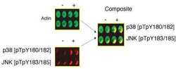
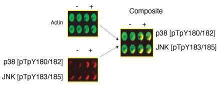
- p38 (pTpY180/182) phosphospecific antibody. RAW cells were untreated (-) or treated (+) with Anisomycin. Two-color In-Cell Western was performed on the LI-COR Odyssey® Infrared Imaging System using actin, p38 (pTpY180/182) (Product # 44-684G), and JNK1/2 (pTpY183/185) (Product # 44-682G) antibodies. Two spectrally distinct dyes were used to detect actin (green, 800 nm channel), p38 (pTpY180/182) or JNK1/2 (pTpY183/185) (red, 700 nm channel), or a composite of both (yellow).
- Submitted by
- Invitrogen Antibodies (provider)
- Main image

- Experimental details
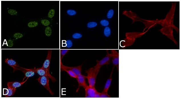
- Immunofluorescent analysis of Phospho-P38 pThr180/pTyr182 Antibody was done on 70% confluent log phase SHSY5Y cells. The cells were fixed with 4% paraformaldehyde for 15 minutes, permeabilized with 0.25% Triton™ X-100 for 10 minutes, and blocked with 5% BSA for 1 hour at room temperature. The cells were labeled with Phospho-P38 pThr180/pTyr182 Antibody (Product # 44-684G) at 1µg/mL in 1% BSA and incubated for 3 hours at room temperature and then labeled with Alexa Fluor 488 Goat Anti-Rabbit IgG Secondary Antibody (Product # A-11008) at a dilution of 1:400 for 45 minutes at room temperature (Panel a: green). Nuclei (Panel b: blue) were stained with SlowFade® Gold Antifade Mountant with DAPI (Product # S36938). F-actin (Panel c: red) was stained with Alexa Fluor 594 Phalloidin (Product # A12381). Panel d is a merged image showing nuclear localization. Panel e is a no primary antibody control. The images were captured at 40X magnification.

- Submitted by
- Invitrogen Antibodies (provider)
- Main image

- Experimental details
- Immunofluorescent analysis of Phospho-P38 pThr180/pTyr182 Antibody was done on 70% confluent log phase SHSY5Y cells. The cells were fixed with 4% paraformaldehyde for 15 minutes, permeabilized with 0.25% Triton™ X-100 for 10 minutes, and blocked with 5% BSA for 1 hour at room temperature. The cells were labeled with Phospho-P38 pThr180/pTyr182 Antibody (Product # 44-684G) at 1µg/mL in 1% BSA and incubated for 3 hours at room temperature and then labeled with Alexa Fluor 488 Goat Anti-Rabbit IgG Secondary Antibody (Product # A-11008) at a dilution of 1:400 for 45 minutes at room temperature (Panel a: green). Nuclei (Panel b: blue) were stained with SlowFade® Gold Antifade Mountant with DAPI (Product # S36938). F-actin (Panel c: red) was stained with Alexa Fluor 594 Phalloidin (Product # A12381). Panel d is a merged image showing nuclear localization. Panel e is a no primary antibody control. The images were captured at 40X magnification.
- Submitted by
- Invitrogen Antibodies (provider)
- Main image

- Experimental details
- p38 (pTpY180/182) phosphospecific antibody. RAW cells were untreated (-) or treated (+) with Anisomycin. Two-color In-Cell Western was performed on the LI-COR Odyssey® Infrared Imaging System using actin, p38 (pTpY180/182) (Product # 44-684G), and JNK1/2 (pTpY183/185) (Product # 44-682G) antibodies. Two spectrally distinct dyes were used to detect actin (green, 800 nm channel), p38 (pTpY180/182) or JNK1/2 (pTpY183/185) (red, 700 nm channel), or a composite of both (yellow).

Supportive validation
- Submitted by
- Invitrogen Antibodies (provider)
- Main image
- Experimental details
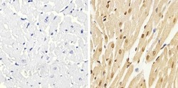
- Immunohistochemistry analysis of p38 (pTpY180/182) showing staining in the cytoplasm and nucleus of paraffin-embedded rat heart tissue (right) compared to a negative control without primary antibody (left). To expose target proteins, antigen retrieval was performed using 10mM sodium citrate (pH 6.0), microwaved for 8-15 min. Following antigen retrieval, tissues were blocked in 3% H2O2-methanol for 15 min at room temperature, washed with ddH2O and PBS, and then probed with a p38 (pTpY180/182) polyclonal antibody (Product # 44-684G) diluted in 3% BSA-PBS at a dilution of 1:20 overnight at 4ºC in a humidified chamber. Tissues were washed extensively in PBST and detection was performed using an HRP-conjugated secondary antibody followed by colorimetric detection using a DAB kit. Tissues were counterstained with hematoxylin and dehydrated with ethanol and xylene to prep for mounting.
- Submitted by
- Invitrogen Antibodies (provider)
- Main image
- Experimental details
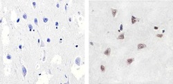
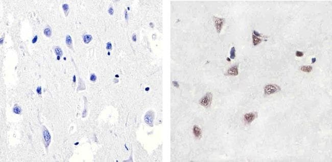
- Immunohistochemistry analysis of p38 (pTpY180/182) showing staining in the cytoplasm and nucleus of paraffin-embedded human brain tissue (right) compared to a negative control without primary antibody (left). To expose target proteins, antigen retrieval was performed using 10mM sodium citrate (pH 6.0), microwaved for 8-15 min. Following antigen retrieval, tissues were blocked in 3% H2O2-methanol for 15 min at room temperature, washed with ddH2O and PBS, and then probed with a p38 (pTpY180/182) polyclonal antibody (Product # 44-684G) diluted in 3% BSA-PBS at a dilution of 1:100 overnight at 4ºC in a humidified chamber. Tissues were washed extensively in PBST and detection was performed using an HRP-conjugated secondary antibody followed by colorimetric detection using a DAB kit. Tissues were counterstained with hematoxylin and dehydrated with ethanol and xylene to prep for mounting.
- Submitted by
- Invitrogen Antibodies (provider)
- Main image

- Experimental details
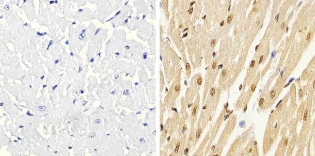
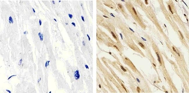
- Immunohistochemistry analysis of p38 (pTpY180/182) showing staining in the cytoplasm and nucleus of paraffin-embedded human heart tissue (right) compared to a negative control without primary antibody (left). To expose target proteins, antigen retrieval was performed using 10mM sodium citrate (pH 6.0), microwaved for 8-15 min. Following antigen retrieval, tissues were blocked in 3% H2O2-methanol for 15 min at room temperature, washed with ddH2O and PBS, and then probed with a p38 (pTpY180/182) polyclonal antibody (Product # 44-684G) diluted in 3% BSA-PBS at a dilution of 1:20 overnight at 4ºC in a humidified chamber. Tissues were washed extensively in PBST and detection was performed using an HRP-conjugated secondary antibody followed by colorimetric detection using a DAB kit. Tissues were counterstained with hematoxylin and dehydrated with ethanol and xylene to prep for mounting.
Supportive validation
- Submitted by
- Invitrogen Antibodies (provider)
- Main image
- Experimental details
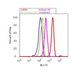
- Flow cytometry analysis of p38 [pTpY180/182] was done on K562 cells treated with anisomycin (25ug/mL, 30 minutes). Cells were fixed with 70% ethanol for 10 minutes, permeabilized with 0.25% Triton™ X-100 for 20 minutes, and blocked with 5% BSA for 30 minutes at room temperature. Cells were labeled with p38 [pTpY180/182] Rabbit Polyclonal Antibody (44684G, red histogram) or with rabbit isotype control (pink histogram) at 3-5 ug/million cells in 2.5% BSA. After incubation at room temperature for 2 hours, the cells were labeled with Alexa Fluor® 488 Goat Anti-Rabbit Secondary Antibody (A11008) at a dilution of 1:400 for 30 minutes at room temperature. The representative 10,000 cells were acquired and analyzed for each sample using an Attune® Acoustic Focusing Cytometer. The purple histogram represents unstained control cells and the green histogram represents no-primary-antibody control.
Supportive validation
- Submitted by
- Invitrogen Antibodies (provider)
- Main image
- Experimental details
- NULL
- Submitted by
- Invitrogen Antibodies (provider)
- Main image
- Experimental details
- NULL
- Submitted by
- Invitrogen Antibodies (provider)
- Main image
- Experimental details
- NULL
- Submitted by
- Invitrogen Antibodies (provider)
- Main image
- Experimental details
- Fig. 6 Effect of candesartan, CoQ10 and their combination on phospho-p38 MAPK expression in rat gastrocnemius. (A) Representative western blots for phospho-p38 MAPK, p38 MAPK, and beta-actin proteins expression (B) Quantification of phospho-p38 MAPK protein expression relative to p38 MAPK (presented as fold change from control). Data are expressed as mean +- S.D (n = 6). a, b, c, d: significantly different from control, tourniquet, candesartan, CoQ10 groups, respectively, at p < 0.05.
- Submitted by
- Invitrogen Antibodies (provider)
- Main image

- Experimental details
- Figure 5 MKP-2 deletion enhances M-CSF-mediated ERK phosphorylation in macrophages. Cells harvested on day 3 of culture, were rendered quiescent in M-CSF free medium then stimulated with recombinant M-CSF (10 ng/mL) for the times indicated. Control cells (0) were left untreated. Whole cell lysates were prepared, and assessed for ( A ) MKP-2 (43 kDa) ( B ) p-ERK (42/44 kDa), ( C ) p-JNK (46/65 kDa), and ( D ) p-p38 (38 kDa) or total controls by Western blotting, as described in the Methods. MKP-2 was not induced in cells that were derived from MKP-2 -/- mice (upper left insert of Panel A). Blots were semi-quantified for fold stimulation by scanning densitometry relative to the background signal. The blot represents the data from 2-3 individual experiments. * p < 0.05, one-tailed unpaired t test comparing MKP-2 -/- to MKP-2 +/+ macrophages.
- Submitted by
- Invitrogen Antibodies (provider)
- Main image
- Experimental details
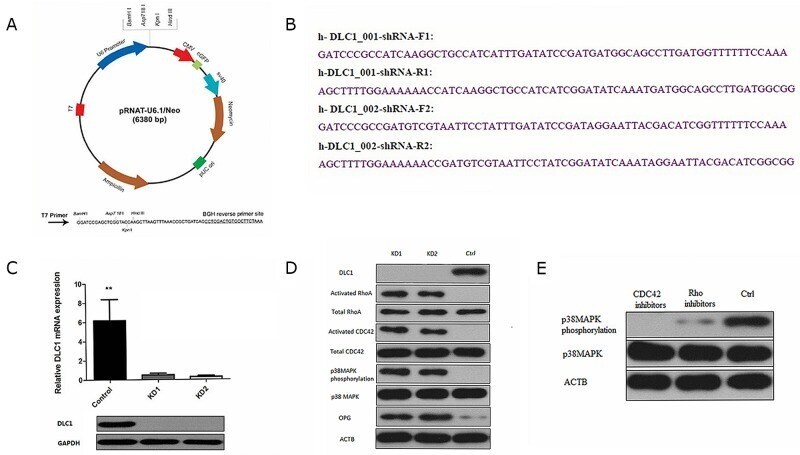
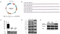
- Figure 6 DLC1 inhibition is critical for mir-141-3p to activate the p38MAPK pathway (A) pRNAT-U6.1/Neo expression vector. (B) Two shRNA constructs and the corresponding primers. (C) Two shRNA constructs stably silence DLC1 in osteoblasts. (D) DLC1 KD activated RhoA and CDC42 and affected p38MAPK phosphorylation. (E) CDC42 inhibition significantly affected p38MAPK phosphorylation.