Explore
 Validate
 Learn
Learn Western blot
Antibody data
- Antibody Data
- Antigen structure
- References [2]
- Comments [0]
- Validations
- Western blot [2]
- Immunohistochemistry [3]
Submit
Validation data
Reference
Comment
Report error
- Product number
- NBP2-24551 - Provider product page

- Provider
- Novus Biologicals
- Product name
- Rabbit Polyclonal c-Maf Antibody
- Antibody type
- Polyclonal
- Description
- Immunogen affinity purified. 100% homologous in human isoforms CRA_a, CRA_b, and CRA_c).
- Reactivity
- Human, Mouse, Rat, Bovine
- Host
- Rabbit
- Isotype
- IgG
- Vial size
- 0.1 mg
- Concentration
- 1.0 mg/ml
- Storage
- Store at 4C short term. Aliquot and store at -20C long term. Avoid freeze-thaw cycles.
Submitted references Disruption of Rest Leads to the Early Onset of Cataracts with the Aberrant Terminal Differentiation of Lens Fiber Cells.
KSHV-encoded miRNAs target MAF to induce endothelial cell reprogramming.
Aoki H, Ogino H, Tomita H, Hara A, Kunisada T
PloS one 2016;11(9):e0163042
PloS one 2016;11(9):e0163042
KSHV-encoded miRNAs target MAF to induce endothelial cell reprogramming.
Hansen A, Henderson S, Lagos D, Nikitenko L, Coulter E, Roberts S, Gratrix F, Plaisance K, Renne R, Bower M, Kellam P, Boshoff C
Genes & development 2010 Jan 15;24(2):195-205
Genes & development 2010 Jan 15;24(2):195-205
No comments: Submit comment
Supportive validation
- Submitted by
- Novus Biologicals (provider)
- Main image
- Experimental details
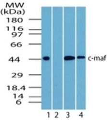
- Western Blot: c-Maf Antibody [NBP2-24551] - Analysis of Lysate from human brain in the 1) absence, 2) presence of immunizing peptide, 3) mouse brain and 4) rat brain probed at 3 ug/ml.

- Submitted by
- Novus Biologicals (provider)
- Main image

- Experimental details
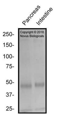
- Western Blot: c-Maf Antibody [NBP2-24551] - Total protein from Human Pancreas and Human Small Intestine was separated on a 7.5% gel by SDS-PAGE, transferred to PVDF membrane and blocked in 5% non-fat milk in TBST. The membrane was probed with 2.0 ug/ml anti-cMaf in 1% block buffer and detected with an anti-rabbit HRP secondary antibody using chemiluminescence.
Supportive validation
- Submitted by
- Novus Biologicals (provider)
- Main image

- Experimental details
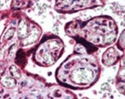
- Immunohistochemistry-Paraffin: c-Maf Antibody [NBP2-24551] - Staining of Human placenta probed with c-maf antibody at 10 ug/ml.
- Submitted by
- Novus Biologicals (provider)
- Main image
- Experimental details
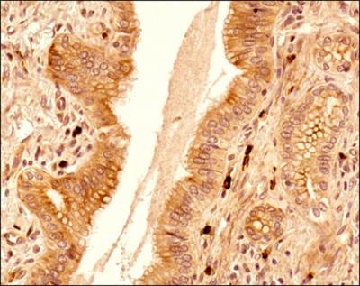
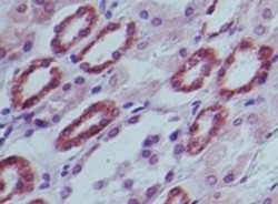
- Immunohistochemistry-Paraffin: c-Maf Antibody [NBP2-24551] - Staining of Human kidney probed with c-maf antibody at 10 ug/ml.

- Submitted by
- Novus Biologicals (provider)
- Main image

- Experimental details
- Immunohistochemistry-Paraffin: c-Maf Antibody [NBP2-24551] - IHC analysis of a formalin fixed paraffin embedded (FFPE) tissue section of human liver cancer using c-MAF antibody at 2ug/ml concentration (1:500 dilution). The primary antibody binding to c-MAF protein was detected using HRP conjugated anti-rabbit secondary antibody with DAB reagent, and the sections were further counterstained with hematoxylin for labeling cellular nuclei. This c-MAF antibody showed a very strong nuclear and moderate to strong staining in the endothelial cells (blood vessels) and in a subset of apparently infiltrating inflammatory cells. Weak to moderate cytoplasmic and nuclear staining was observed in liver cancer cells and the cells of tumor stroma including fibroblasts.