Explore
 Validate
 Learn
Learn Western blot
Antibody data
- Antibody Data
- Antigen structure
- References [0]
- Comments [0]
- Validations
- Western blot [1]
- Immunocytochemistry [1]
Submit
Validation data
Reference
Comment
Report error
- Product number
- MAB8227 - Provider product page

- Provider
- R&D Systems
- Product name
- Human c-Maf Antibody
- Antibody type
- Monoclonal
- Description
- Protein A or G purified from hybridoma culture supernatant. Detects human c-Maf in direct ELISA and Western Blot.
- Reactivity
- Human
- Host
- Mouse
- Conjugate
- Unconjugated
- Antigen sequence
O75444- Isotype
- IgG
- Antibody clone number
- 883926
- Vial size
- 100 ug
- Concentration
- LYOPH
- Storage
- Use a manual defrost freezer and avoid repeated freeze-thaw cycles. 12 months from date of receipt, -20 to -70 °C as supplied. 1 month, 2 to 8 °C under sterile conditions after reconstitution. 6 months, -20 to -70 °C under sterile conditions after reconstitution.
No comments: Submit comment
Supportive validation
- Submitted by
- R&D Systems (provider)
- Main image
- Experimental details
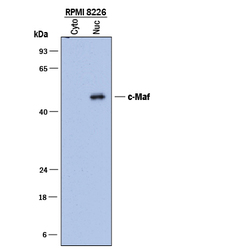
- Detection of Human c-Maf by Western Blot. Western blot shows lysates of RPMI 8226 human multiple myeloma cell line. Gels were loaded with 40 μg of cytoplasmic (Cyto) and 20 μg of nuclear (Nuc) extracts. PVDF membrane was probed with 2 µg/mL of Mouse Anti-Human c-Maf Monoclonal Antibody (Catalog # MAB8227) followed by HRP-conjugated Anti-Mouse IgG Secondary Antibody (Catalog # HAF018). A specific band was detected for c-Maf at approximately 48 kDa (as indicated). This experiment was conducted under reducing conditions and using Immunoblot Buffer Group 1.

Supportive validation
- Submitted by
- R&D Systems (provider)
- Main image
- Experimental details
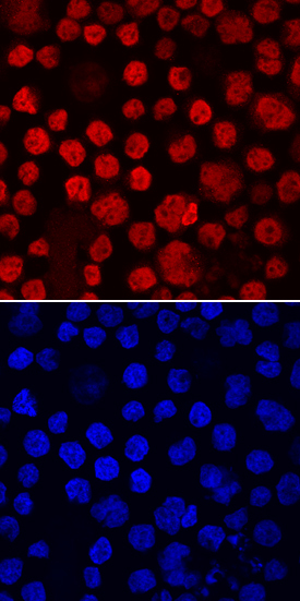
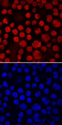
- c-Maf in RPMI 8226 Human Cell Line. c-Maf was detected in immersion fixed RPMI 8226 human multiple myeloma cell line using Mouse Anti-Human c-Maf Monoclonal Antibody (Catalog # MAB8227) at 10 µg/mL for 3 hours at room temperature. Cells were stained using the NorthernLights™ 557-conjugated Anti-Mouse IgG Secondary Antibody (red, upper panel; Catalog # NL007) and counterstained with DAPI (blue, lower panel). Specific staining was localized to nuclei. View our protocol for Fluorescent ICC Staining of Non-adherent Cells.