Explore
 Validate
 Learn
Learn Flow cytometry
Antibody data
- Antibody Data
- Antigen structure
- References [8]
- Comments [0]
- Validations
- Flow cytometry [1]
- Other assay [2]
Submit
Validation data
Reference
Comment
Report error
- Product number
- 12-9855-41 - Provider product page

- Provider
- Invitrogen Antibodies
- Product name
- c-MAF Monoclonal Antibody (sym0F1), PE, eBioscience™
- Antibody type
- Monoclonal
- Antigen
- Other
- Description
- Description: The sym0F1 monoclonal antibody reacts with human and mouse c-Maf, a 42 kDa basic leucine zipper transcription factor shown to be involved in the neural, ocular and hematopoietic systems. In hematopoietic cells, it was first shown to be crucial for IL-4 expression in Th2 and was the first transcription factor believed to be Th subset-specific. More recent evidence shows that specific phospho-tyrosine residues lead to upregulation of IL-4. c-Maf has also been shown to be important to differentiation and function in both Th17 and Tfh cells. It drives expression of IL-21 in both cell types, while promoting expression of IL-23R in Th17 and CXCR5 in Tfh as well. Sym0F1 does not cross-react with MafA or MafB. Applications Reported: This sym0F1 antibody has been reported for use in intracellular staining followed by flow cytometric analysis. Applications Tested: This sym0F1 antibody has been pre-titrated and tested by intracellular staining followed by flow cytometric analysis of Th17-polarized mouse splenocytes using the Foxp3/Transcription Factor Staining Buffer Set (Product # 00-5523-00) and protocol. Please refer to Best Protocols: Protocol B: One step protocol for (nuclear) intracellular proteins located under the Resources Tab online. This can be used at 5 µL (0.06 µg) per test. A test is defined as the amount (µg) of antibody that will stain a cell sample in a final volume of 100 µL. Cell number should be determined empirically but can range from 10^5 to 10^8 cells/test. Excitation: 488-561 nm; Emission: 578 nm; Laser: Blue Laser, Green Laser, Yellow-Green Laser. Filtration: 0.2 µm post-manufacturing filtered.
- Reactivity
- Human, Mouse
- Host
- Mouse
- Conjugate
- Yellow dye
- Isotype
- IgG
- Antibody clone number
- sym0F1
- Vial size
- 25 Tests
- Concentration
- 5 µL/Test
- Storage
- 4° C, store in dark, DO NOT FREEZE!
Submitted references Natural γδT17 cell development and functional acquisition is governed by the mTORC2-c-Maf-controlled mitochondrial fission pathway.
GM-CSF-activated human dendritic cells promote type 1 T follicular helper cell polarization in a CD40-dependent manner.
Oncogenic Vav1-Myo1f induces therapeutically targetable macrophage-rich tumor microenvironment in peripheral T cell lymphoma.
Transcriptome dynamics of CD4(+) T cells during malaria maps gradual transit from effector to memory.
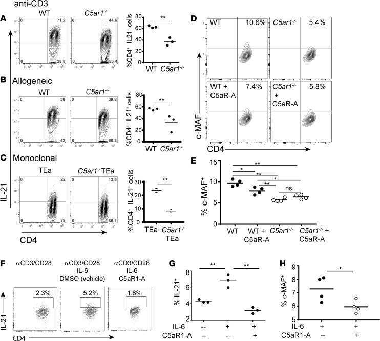
C5aR1 regulates T follicular helper differentiation and chronic graft-versus-host disease bronchiolitis obliterans.
Epidermal Growth Factor Receptor Expression Licenses Type-2 Helper T Cells to Function in a T Cell Receptor-Independent Fashion.
Characterization of c-Maf(+)Foxp3(-) Regulatory T Cells Induced by Repeated Stimulation of Antigen-Presenting B Cells.
Human circulating PD-1+CXCR3-CXCR5+ memory Tfh cells are highly functional and correlate with broadly neutralizing HIV antibody responses.
Wang Y, Qin H, Cai Y, Chen X, Li H, Montoya-Durango DE, Ding C, Hu X, Chariker JH, Sarojini H, Chien S, Rouchka EC, Zhang HG, Zheng J, Qiu F, Yan J
iScience 2023 May 19;26(5):106630
iScience 2023 May 19;26(5):106630
GM-CSF-activated human dendritic cells promote type 1 T follicular helper cell polarization in a CD40-dependent manner.
Korniotis S, Saichi M, Trichot C, Hoffmann C, Amblard E, Viguier A, Grondin S, Noel F, Mattoo H, Soumelis V
Journal of cell science 2022 Nov 1;135(21)
Journal of cell science 2022 Nov 1;135(21)
Oncogenic Vav1-Myo1f induces therapeutically targetable macrophage-rich tumor microenvironment in peripheral T cell lymphoma.
Cortes JR, Filip I, Albero R, Patiño-Galindo JA, Quinn SA, Lin WW, Laurent AP, Shih BB, Brown JA, Cooke AJ, Mackey A, Einson J, Zairis S, Rivas-Delgado A, Laginestra MA, Pileri S, Campo E, Bhagat G, Ferrando AA, Rabadan R, Palomero T
Cell reports 2022 Apr 19;39(3):110695
Cell reports 2022 Apr 19;39(3):110695
Transcriptome dynamics of CD4(+) T cells during malaria maps gradual transit from effector to memory.
Soon MSF, Lee HJ, Engel JA, Straube J, Thomas BS, Pernold CPS, Clarke LS, Laohamonthonkul P, Haldar RN, Williams CG, Lansink LIM, Moreira ML, Bramhall M, Koufariotis LT, Wood S, Chen X, James KR, Lönnberg T, Lane SW, Belz GT, Engwerda CR, Khoury DS, Davenport MP, Svensson V, Teichmann SA, Haque A
Nature immunology 2020 Dec;21(12):1597-1610
Nature immunology 2020 Dec;21(12):1597-1610
C5aR1 regulates T follicular helper differentiation and chronic graft-versus-host disease bronchiolitis obliterans.
Verghese DA, Chun N, Paz K, Fribourg M, Woodruff TM, Flynn R, Hu Y, Xiong H, Zhang W, Yi Z, Du J, Blazar BR, Heeger PS
JCI insight 2018 Dec 20;3(24)
JCI insight 2018 Dec 20;3(24)
Epidermal Growth Factor Receptor Expression Licenses Type-2 Helper T Cells to Function in a T Cell Receptor-Independent Fashion.
Minutti CM, Drube S, Blair N, Schwartz C, McCrae JC, McKenzie AN, Kamradt T, Mokry M, Coffer PJ, Sibilia M, Sijts AJ, Fallon PG, Maizels RM, Zaiss DM
Immunity 2017 Oct 17;47(4):710-722.e6
Immunity 2017 Oct 17;47(4):710-722.e6
Characterization of c-Maf(+)Foxp3(-) Regulatory T Cells Induced by Repeated Stimulation of Antigen-Presenting B Cells.
Chien CH, Yu HC, Chen SY, Chiang BL
Scientific reports 2017 Apr 12;7:46348
Scientific reports 2017 Apr 12;7:46348
Human circulating PD-1+CXCR3-CXCR5+ memory Tfh cells are highly functional and correlate with broadly neutralizing HIV antibody responses.
Locci M, Havenar-Daughton C, Landais E, Wu J, Kroenke MA, Arlehamn CL, Su LF, Cubas R, Davis MM, Sette A, Haddad EK, International AIDS Vaccine Initiative Protocol C Principal Investigators, Poignard P, Crotty S
Immunity 2013 Oct 17;39(4):758-69
Immunity 2013 Oct 17;39(4):758-69
No comments: Submit comment
Supportive validation
- Submitted by
- Invitrogen Antibodies (provider)
- Main image
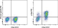
- Experimental details
- Staining of 3-day Th17-polarized mouse splenocytes with Anti-Mouse CD4 FITC (Product # 11-0042-82) and Mouse IgG2b K Isotype Control PE (Product # 12-4732-81) (left) or Anti-Human/Mouse c-Maf PE (right). Viable cells in the lymphocyte gate, as determined by Fixable Viability Dye eFluor® 506 (Product # 65-0866-14), were used for analysis.
- Conjugate
- Yellow dye

Supportive validation
- Submitted by
- Invitrogen Antibodies (provider)
- Main image

- Experimental details
- NULL
- Conjugate
- Yellow dye

- Submitted by
- Invitrogen Antibodies (provider)
- Main image

- Experimental details
- NULL
- Conjugate
- Yellow dye