Explore
 Validate
 Learn
Learn Flow cytometry
Antibody data
- Antibody Data
- Antigen structure
- References [7]
- Comments [0]
- Validations
- Flow cytometry [1]
- Other assay [2]
Submit
Validation data
Reference
Comment
Report error
- Product number
- 50-9855-82 - Provider product page

- Provider
- Invitrogen Antibodies
- Product name
- c-MAF Monoclonal Antibody (sym0F1), eFluor™ 660, eBioscience™
- Antibody type
- Monoclonal
- Antigen
- Other
- Description
- Description: The sym0F1 monoclonal antibody reacts with human and mouse c-Maf, a 42 kDa basic leucine zipper transcription factor shown to be involved in the neural, ocular and hematopoietic systems. In hematopoietic cells, it was first shown to be crucial for IL-4 expression in Th2 and was the first transcription factor believed to be Th subset-specific. More recent evidence shows that specific phospho-tyrosine residues lead to upregulation of IL-4. c-Maf has also been shown to be important to differentiation and function in both Th17 and Tfh cells. It drives expression of IL-21 in both cell types, while promoting expression of IL-23R in Th17 and CXCR5 in Tfh as well.
- Antibody clone number
- sym0F1
- Concentration
- 0.2 mg/mL
Submitted references An immunoregulatory and tissue-residency program modulated by c-MAF in human T(H)17 cells.
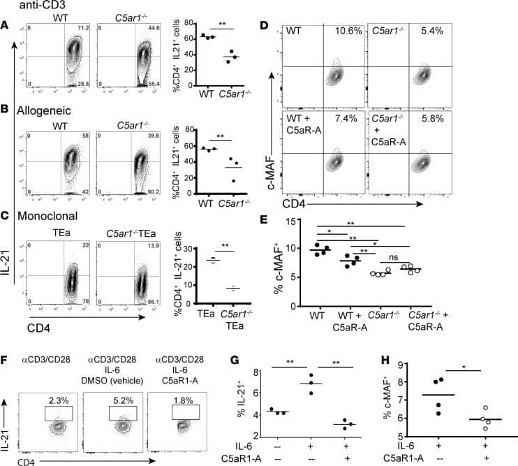
C5aR1 regulates T follicular helper differentiation and chronic graft-versus-host disease bronchiolitis obliterans.
Cytomegalovirus-Specific IL-10-Producing CD4+ T Cells Are Governed by Type-I IFN-Induced IL-27 and Promote Virus Persistence.
Human circulating PD-1+CXCR3-CXCR5+ memory Tfh cells are highly functional and correlate with broadly neutralizing HIV antibody responses.
Tyrosine phosphorylation of c-Maf enhances the expression of IL-4 gene.
Bcl6 and Maf cooperate to instruct human follicular helper CD4 T cell differentiation.
The proto-oncogene c-maf is responsible for tissue-specific expression of interleukin-4.
Aschenbrenner D, Foglierini M, Jarrossay D, Hu D, Weiner HL, Kuchroo VK, Lanzavecchia A, Notarbartolo S, Sallusto F
Nature immunology 2018 Oct;19(10):1126-1136
Nature immunology 2018 Oct;19(10):1126-1136
C5aR1 regulates T follicular helper differentiation and chronic graft-versus-host disease bronchiolitis obliterans.
Verghese DA, Chun N, Paz K, Fribourg M, Woodruff TM, Flynn R, Hu Y, Xiong H, Zhang W, Yi Z, Du J, Blazar BR, Heeger PS
JCI insight 2018 Dec 20;3(24)
JCI insight 2018 Dec 20;3(24)
Cytomegalovirus-Specific IL-10-Producing CD4+ T Cells Are Governed by Type-I IFN-Induced IL-27 and Promote Virus Persistence.
Clement M, Marsden M, Stacey MA, Abdul-Karim J, Gimeno Brias S, Costa Bento D, Scurr MJ, Ghazal P, Weaver CT, Carlesso G, Clare S, Jones SA, Godkin A, Jones GW, Humphreys IR
PLoS pathogens 2016 Dec;12(12):e1006050
PLoS pathogens 2016 Dec;12(12):e1006050
Human circulating PD-1+CXCR3-CXCR5+ memory Tfh cells are highly functional and correlate with broadly neutralizing HIV antibody responses.
Locci M, Havenar-Daughton C, Landais E, Wu J, Kroenke MA, Arlehamn CL, Su LF, Cubas R, Davis MM, Sette A, Haddad EK, International AIDS Vaccine Initiative Protocol C Principal Investigators, Poignard P, Crotty S
Immunity 2013 Oct 17;39(4):758-69
Immunity 2013 Oct 17;39(4):758-69
Tyrosine phosphorylation of c-Maf enhances the expression of IL-4 gene.
Lai CY, Lin SY, Wu CK, Yeh LT, Sytwu HK, Miaw SC
Journal of immunology (Baltimore, Md. : 1950) 2012 Aug 15;189(4):1545-50
Journal of immunology (Baltimore, Md. : 1950) 2012 Aug 15;189(4):1545-50
Bcl6 and Maf cooperate to instruct human follicular helper CD4 T cell differentiation.
Kroenke MA, Eto D, Locci M, Cho M, Davidson T, Haddad EK, Crotty S
Journal of immunology (Baltimore, Md. : 1950) 2012 Apr 15;188(8):3734-44
Journal of immunology (Baltimore, Md. : 1950) 2012 Apr 15;188(8):3734-44
The proto-oncogene c-maf is responsible for tissue-specific expression of interleukin-4.
Ho IC, Hodge MR, Rooney JW, Glimcher LH
Cell 1996 Jun 28;85(7):973-83
Cell 1996 Jun 28;85(7):973-83
No comments: Submit comment
Supportive validation
- Submitted by
- Invitrogen Antibodies (provider)
- Main image
- Experimental details
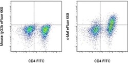
- Intracellular staining of 3-day Th17-polarized mouse splenocytes with Anti-Mouse CD4 FITC (Product # 11-0042-82) and 0.06 µg of Mouse IgG2b K Isotype Control eFluor® 660 (Product # 50-4732-82) (left) or 0.06 µg of Anti-Human/Mouse c-Maf eFluor® 660 (right) using the Foxp3/Transcription Factor Buffer Set (Product # 00-5523-00). Cells in the lymphocyte gate were used for analysis.

Supportive validation
- Submitted by
- Invitrogen Antibodies (provider)
- Main image
- Experimental details
- NULL

- Submitted by
- Invitrogen Antibodies (provider)
- Main image

- Experimental details
- NULL