Explore
 Validate
 Learn
Learn Western blot
 Immunoprecipitation
Antibody data
- Antibody Data
- Antigen structure
- References [0]
- Comments [0]
- Validations
- Western blot [2]
- Immunocytochemistry [2]
- Immunohistochemistry [2]
- Other assay [1]
Submit
Validation data
Reference
Comment
Report error
- Product number
- A700-045 - Provider product page

- Provider
- Invitrogen Antibodies
- Product name
- c-Maf Recombinant Rabbit Monoclonal Antibody (BLR045F)
- Antibody type
- Monoclonal
- Antigen
- Other
- Reactivity
- Human, Mouse
- Host
- Rabbit
- Isotype
- IgG
- Antibody clone number
- BLR045F
- Vial size
- 100 µL
- Concentration
- 1000 µg/mL
- Storage
- 4° C
No comments: Submit comment
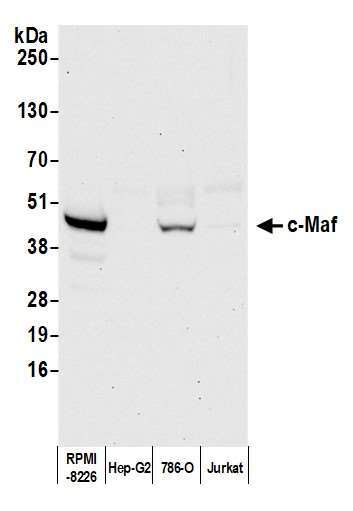
Supportive validation
- Submitted by
- Invitrogen Antibodies (provider)
- Main image
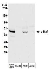
- Experimental details
- Detection of human c-Maf by western blot. Samples: Whole cell lysate (50 µg) from RPMI-8226, Hep-G2, 786-O, and Jurkat cells prepared using NETN lysis buffer. Antibody: Rabbit anti-c-Maf recombinant monoclonal antibody [BL-664A-5F2] (Product # A700-045 lot 3) used at 1:1000. Secondary: HRP-conjugated goat anti-rabbit IgG (A120-101P). Chemiluminescence with an exposure time of 30 seconds.
- Submitted by
- Invitrogen Antibodies (provider)
- Main image
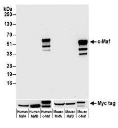
- Experimental details
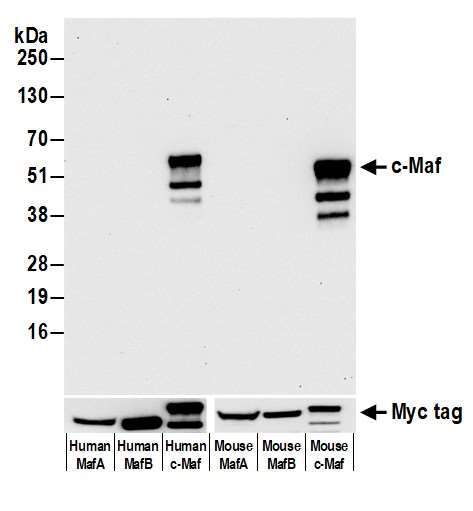
- Detection of human c-Maf by western blot of HEK293T transfected with myc tagged human or mouse MafA, MafB, or c-Maf. Antibody: Rabbit anti-c-Maf recombinant monoclonal antibody [BLR045R] (Product # A700-045 lot 3) used at 1:1000. Secondary: HRP-conjugated goat anti-rabbit IgG (A120-101P). Chemiluminescence with an exposure time of 30 seconds. Lower Panel: Rabbit anti-Myc Tag recombinant monoclonal antibody [BLRE01G] (Product # A191-101A).
Supportive validation
- Submitted by
- Invitrogen Antibodies (provider)
- Main image
- Experimental details
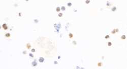
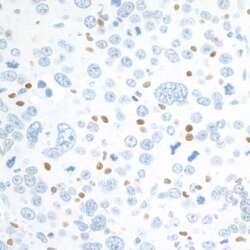
- Detection of human c-MAF by immunocytochemistry.Sample: FFPE section of RPMI-8226 cells.Antibody: Rabbit anti-c-MAF recombinant monoclonal antibody [BLR045F] (Product # A700-045; lot 3) used at 1:125.Secondary: HRP-conjugated goat anti-rabbit IgG (Product # A120-501P). Substrate: DAB.
- Submitted by
- Invitrogen Antibodies (provider)
- Main image
- Experimental details
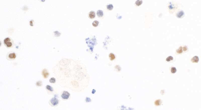
- Detection of human c-MAF by immunocytochemistry.Sample: FFPE section of RPMI-8226 cells.Antibody: Rabbit anti-c-MAF recombinant monoclonal antibody [BLR045F] (Product # A700-045; lot 3) used at 1:125.Secondary: HRP-conjugated goat anti-rabbit IgG (Product # A120-501P). Substrate: DAB.

Supportive validation
- Submitted by
- Invitrogen Antibodies (provider)
- Main image

- Experimental details
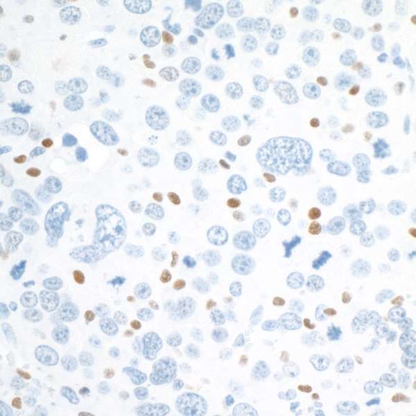
- Detection of human c-MAF by immunohistochemistry.Sample: FFPE section of human tonsil.Antibody: Rabbit anti-c-MAF recombinant monoclonal antibody [BLR045F] (Product # A700-045 lot 3) used at 1:125.Secondary: HRP-conjugated goat anti-rabbit IgG (A120-501P). Substrate: DAB.
- Submitted by
- Invitrogen Antibodies (provider)
- Main image
- Experimental details
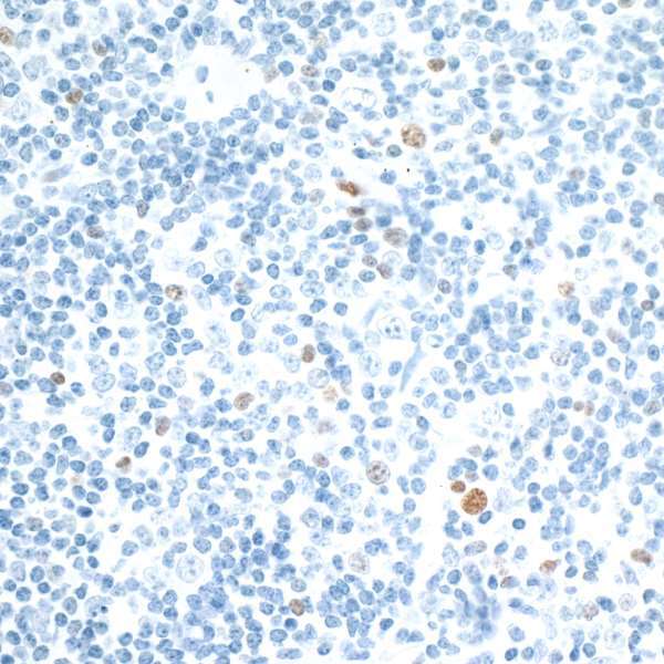
- Detection of mouse c-MAF by immunohistochemistry.Sample: FFPE section of mouse renal cell carcinoma.Antibody: Rabbit anti-c-MAF recombinant monoclonal antibody [BLR045F] (Product # A700-045 lot 3) used at 1:125.Secondary: HRP-conjugated goat anti-rabbit IgG (A120-501P). Substrate: DAB.
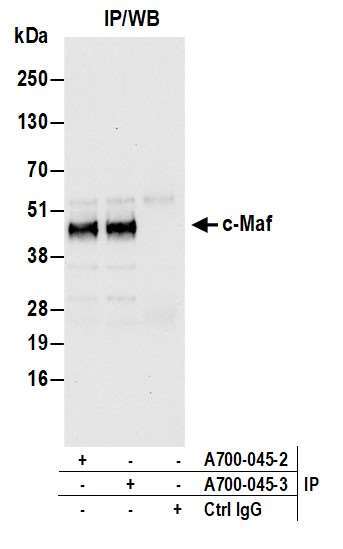
Supportive validation
- Submitted by
- Invitrogen Antibodies (provider)
- Main image
- Experimental details
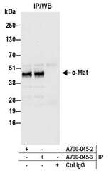
- Detection of human c-Maf by western blot of immunoprecipitates. Samples: Whole cell lysate (1.0 mg per IP reaction; 20% of IP loaded) from 293T cells prepared using NETN lysis buffer. Antibodies: Rabbit anti-c-Maf recombinant monoclonal antibody [BL-664A-5F2] (Product # A700-045 lot 3) used for IP at 6 µL per reaction. c-Maf was also immunoprecipitated by a previous lot of this antibody (lot 2). For blotting immunoprecipitated c-Maf (Product # A700-045) was used at 1:1000. Chemiluminescence with an exposure time of 10 seconds.