Explore
 Validate
 Learn
Learn Western blot
 ELISA
ELISAAntibody data
- Antibody Data
- Antigen structure
- References [0]
- Comments [0]
- Validations
- Western blot [2]
- Immunocytochemistry [1]
Submit
Validation data
Reference
Comment
Report error
- Product number
- AM32520PU-N - Provider product page

- Provider
- Acris Antibodies GmbH
- Product name
- anti MAPKAP Kinase-2 pThr334
- Antibody type
- Monoclonal
- Antigen
- Synthetic peptide containing the sequence and surrounding amino acids phosphorylated Thr334 of Human, Mouse, Rat, Bovine and Chicken MAPKAP Kinase 2. Immunizing peptide shared 66% homology with phospho-MAPKAP-K3 at Thr313
- Reactivity
- Human, Mouse, Rat
- Host
- Mouse
- Isotype
- IgG
- Antibody clone number
- 9F10.2
- Vial size
- 0.1 mg
- Concentration
- 1.0 mg/ml
No comments: Submit comment
Supportive validation
- Submitted by
- Acris Antibodies GmbH (provider)
- Main image

- Experimental details
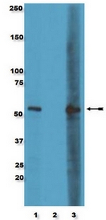
- Western Blot Analysis: PC12 cell lysates were resolved by electrophoresis, transferred to PVDF, and probed with were either unblocked (Lane 1), pre-blocked with phosphorylated peptide (Lane 2), or pre-blocked with unphosphorylated peptide (Lane 3) Anti-phospho-MAPKAPK2 (Thr334), clone 9F10.2 (1/1000). Proteins were visualized using a goat anti-mouse secondary antibody conjugated to HRP and a chemiluminescence detection system. Arrow indicates phosphorylated MAPKAPK2 (~50 kDa)
- Submitted by
- Acris Antibodies GmbH (provider)
- Main image
- Experimental details
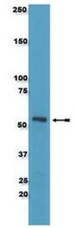
- Western Blot Analysis:Â PC12 cell lysate was resolved by SDS-PAGE, transferred to PVDF, and probed with anti-phospho-MAPKAPK2 (Thr334), clone 9F10.2 (1/1,000). Proteins were visualized using a goat anti-mouse secondary antibody conjugated to HRP and a chemiluminescence detection system. Arrow indicates phosphorylated MAPKAPK2 (~50 kDa).
Supportive validation
- Submitted by
- Acris Antibodies GmbH (provider)
- Main image

- Experimental details
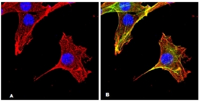
- Immunofluorescence Confocal Microscopy Analysis: NIH/3T3 cells were fixed, permeabilized, and stained using Anti-phospho-MAPKAPK2 (Thr334), clone 9F10.2 (Red). Cells were costained for actin and nuclear staining with Phalloidin, AlexaFluor® 488 conjugate (Green) and DAPI (Blue), respectively.