Explore
 Validate
 Learn
Learn Western blot
 Immunocytochemistry
Antibody data
- Antibody Data
- Antigen structure
- References [4]
- Comments [0]
- Validations
- Immunocytochemistry [2]
- Immunohistochemistry [2]
- Other assay [3]
Submit
Validation data
Reference
Comment
Report error
- Product number
- PA5-20811 - Provider product page

- Provider
- Invitrogen Antibodies
- Product name
- RIP1 Polyclonal Antibody
- Antibody type
- Polyclonal
- Antigen
- Synthetic peptide
- Description
- A suggested positive control is rat kidney tissue lysate. PA5-20811 can be used with blocking peptide PEP-0925.
- Reactivity
- Human, Mouse, Rat
- Host
- Rabbit
- Isotype
- IgG
- Vial size
- 100 μg
- Concentration
- 1.0 mg/mL
- Storage
- Maintain refrigerated at 2-8°C for up to 3 months. For long term storage store at -20°C
Submitted references Coenzyme Q10 encapsulated in micelles ameliorates osteoarthritis by inhibiting inflammatory cell death.
Mitochondrial Transplantation Ameliorates the Development and Progression of Osteoarthritis.
Fractalkine Is Linked to the Necrosome Pathway in Acute Pulmonary Inflammation.
Would Colloidal Gold Nanocarriers Present An Effective Diagnosis Or Treatment For Ischemic Stroke?
Na HS, Woo JS, Kim JH, Lee JS, Um IG, Cho KH, Kim GH, Cho ML, Chung SJ, Park SH
PloS one 2022;17(6):e0270351
PloS one 2022;17(6):e0270351
Mitochondrial Transplantation Ameliorates the Development and Progression of Osteoarthritis.
Lee AR, Woo JS, Lee SY, Na HS, Cho KH, Lee YS, Lee JS, Kim SA, Park SH, Kim SJ, Cho ML
Immune network 2022 Apr;22(2):e14
Immune network 2022 Apr;22(2):e14
Fractalkine Is Linked to the Necrosome Pathway in Acute Pulmonary Inflammation.
Ngamsri KC, Gamper-Tsigaras J, Reutershan J, Konrad FM
Frontiers in medicine 2021;8:591790
Frontiers in medicine 2021;8:591790
Would Colloidal Gold Nanocarriers Present An Effective Diagnosis Or Treatment For Ischemic Stroke?
Amani H, Mostafavi E, Alebouyeh MR, Arzaghi H, Akbarzadeh A, Pazoki-Toroudi H, Webster TJ
International journal of nanomedicine 2019;14:8013-8031
International journal of nanomedicine 2019;14:8013-8031
No comments: Submit comment
Supportive validation
- Submitted by
- Invitrogen Antibodies (provider)
- Main image

- Experimental details
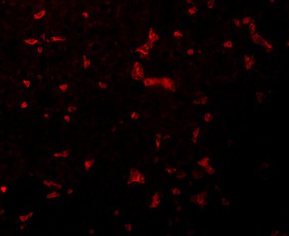
- Immunofluorescence of RIPK1 in Mouse Kidney cells with RIP1 Polyclonal Antibody (Product # PA5-20811) at 20 µg/mL.

- Submitted by
- Invitrogen Antibodies (provider)
- Main image

- Experimental details
- Immunofluorescence of RIPK1 in Mouse Kidney cells with RIP1 Polyclonal Antibody (Product # PA5-20811) at 20 µg/mL.
Supportive validation
- Submitted by
- Invitrogen Antibodies (provider)
- Main image
- Experimental details
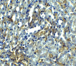
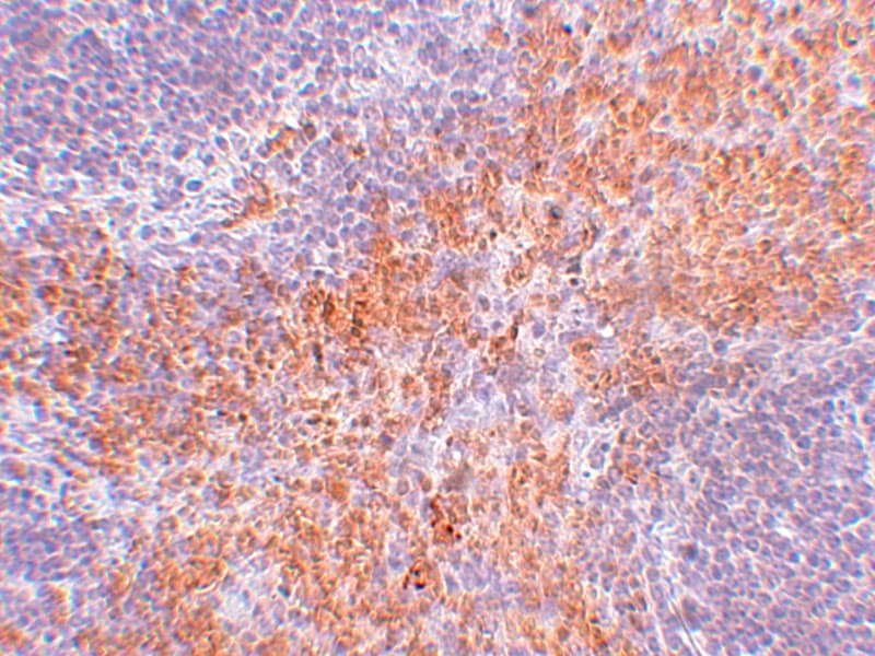
- Immunohistochemistry of RIPK1 in mouse kidney tissue with RIP1 Polyclonal Antibody (Product # PA5-20811) at 2.5 µg/mL.

- Submitted by
- Invitrogen Antibodies (provider)
- Main image
- Experimental details
- Immunohistochemistry of RIPK1 in mouse kidney tissue with RIP1 Polyclonal Antibody (Product # PA5-20811) at 2.5 µg/mL.
Supportive validation
- Submitted by
- Invitrogen Antibodies (provider)
- Main image

- Experimental details
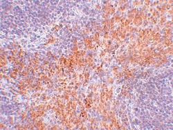
- Figure 4 In vivo detection of necrosome activation after the onset of inflammation. Expression of the necrosome-related receptor-interacting serine/threonine-protein kinase (RIPK)1 and RIPK3 at the gene (A) ( n = 8) and protein (B) levels was detected in the lungs of C57BL/6J and CX 3 CR1 -/- mice 24 h after LPS exposure. The protein expression was further analyzed by measuring the phosphorylation and therefore activation of RIPK1, RIPK3, and mixed lineage kinase domain-like protein (MLKL). Representative blots are shown, and the density was assessed ( n = 3-4). The intensity of the blots was evaluated by ImageJ. (C) The phosphorylated form of MLKL was also detected by immunofluorescence in the lungs of C57BL/6J and CX 3 CR1 -/- mice (pMLKL appears green; original magnification 63x; one representative image of six is shown; n = 4). (D) Gene expression of IL-33 was determined in the lung tissue of C57BL/6J and CX 3 CR1 -/- mice 24 h after LPS exposure ( n = 6-8). The release of IL-33 and HMGB1 was evaluated in the bronchoalveolar lavage of C57BL/6J and CX 3 CR1 -/- mice ( n = 6-8). (E) Schematic diagram of LPS-dependent necrosome formation in murine lungs. Transmembrane toll-like receptor (TLR) 4 activates (toll/interleukin-1 receptor) domain-containing adaptor protein (TRIF), leading to the phosphorylation of RIPK1 and RIPK3. Necrosome activation induces the phosphorylation of MLKL, resulting in necroptosis and the release of damage-associated patterns (DAMPs). All the data a

- Submitted by
- Invitrogen Antibodies (provider)
- Main image
- Experimental details
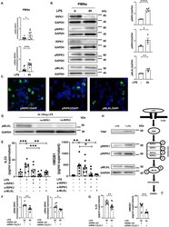
- Figure 7 In vitro necrosome activation on human PMNs. (A) Necrosome-related protein kinase RIPK1 and RIPK3 expression in human PMNs after LPS stimulation ( n = 8) was detected by RT-PCR. (B) Immunoblots of RIPK1, RIPK3, MLKL and their phosphorylated forms were obtained from cell lysates of human PMNs after LPS stimulation (representative blots of n = 4-5 are shown). The intensity of the blots was evaluated by ImageJ. (C) Phosphorylation of RIPK1, RIPK3, and MLKL was detected in human PMNs after LPS (original magnification 63x; one representative image of five is shown; n = 4). (D) Effects of specific RIPK1 and RIPK3 inhibition on MLKL phosphorylation in human PMNs after LPS exposure were evaluated (representative blots from 3 independent experiments; n = 3). (E) Human IL-33 and HMGB1 were determined in the supernatant of stimulated and treated PMNs as indicated ( n = 6-8). (F) Effects of fractalkine depletion on necrosome-related RIPK1 and RIPK3 expression in human PMNs ( n = 6-8) and on the release of human alarmins IL-33 and HMGB1 (G) were evaluated ( n = 8-12). All the data are presented as the mean +- SEM; * p < 0.05; ** p < 0.01; *** p < 0.001; Student''s t -test to compare two groups or one-way ANOVA + Bonferroni test to compare multiple groups. (H) Schematic diagram of LPS-dependent necrosome formation in human PMNs. Transmembrane toll-like receptor (TLR) 4 activates (toll/interleukin-1 receptor) domain-containing adaptor protein (TRIF), leading to the phosphorylation

- Submitted by
- Invitrogen Antibodies (provider)
- Main image
- Experimental details
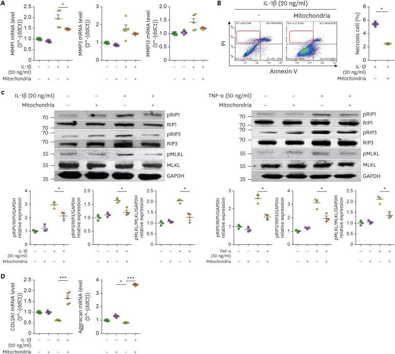
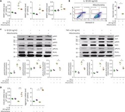
- Expression of cell death and cartilage regulatory markers following mitochondria administration. Human OA chondrocytes were cultured in the presence or absence of IL-1beta (20 ng/mL) or TNF-alpha (50 ng/ml) and mitochondria (5 mug) for 24 h. (A) mRNA levels of catabolic factors were analyzed by qPCR. (B) Necrosis analysis by annexinV/PI staining. (C) Expression of phospho- or total of RIPK1, RIPK3, MLKL in OA chondrocytes was analyzed by western blotting. (D) mRNA levels of chondrogenesis markers in OA chondrocytes. Data are means +- SEM. * p