Explore
 Validate
 Learn
Learn Western blot
 Immunohistochemistry
Antibody data
- Antibody Data
- Antigen structure
- References [3]
- Comments [0]
- Validations
- Immunohistochemistry [1]
- Blocking/Neutralizing [1]
Submit
Validation data
Reference
Comment
Report error
- Product number
- AF-237-NA - Provider product page

- Provider
- R&D Systems
- Product name
- Human FGF-5 Antibody
- Antibody type
- Polyclonal
- Description
- Immunogen affinity purified. Detects human FGF-5 in direct ELISAs and Western blots. In direct ELISAs, less than 1% cross-reactivity with recombinant human (rh) FGF-4, rhFGF-6, rhFGF-7, recombinant mouse FGF-8b, rhFGF-9, rhFGF acidic, and rhFGF basic is observed.
- Reactivity
- Human
- Host
- Goat
- Conjugate
- Unconjugated
- Antigen sequence
Q8NF90- Isotype
- IgG
- Vial size
- 100 ug
- Concentration
- LYOPH
- Storage
- Use a manual defrost freezer and avoid repeated freeze-thaw cycles. 12 months from date of receipt, -20 to -70 °C as supplied. 1 month, 2 to 8 °C under sterile conditions after reconstitution. 6 months, -20 to -70 °C under sterile conditions after reconstitution.
Submitted references Direct Conversion of Mouse Fibroblasts into Neural Stem Cells by Chemical Cocktail Requires Stepwise Activation of Growth Factors and Nup210.
Fibroblast growth factor receptors as therapeutic targets in human melanoma: synergism with BRAF inhibition.
FGF5 as an oncogenic factor in human glioblastoma multiforme: autocrine and paracrine activities.
Tang Y, Xiong S, Yu P, Liu F, Cheng L
Cell reports 2018 Jul 31;24(5):1355-1362.e3
Cell reports 2018 Jul 31;24(5):1355-1362.e3
Fibroblast growth factor receptors as therapeutic targets in human melanoma: synergism with BRAF inhibition.
Metzner T, Bedeir A, Held G, Peter-Vörösmarty B, Ghassemi S, Heinzle C, Spiegl-Kreinecker S, Marian B, Holzmann K, Grasl-Kraupp B, Pirker C, Micksche M, Berger W, Heffeter P, Grusch M
The Journal of investigative dermatology 2011 Oct;131(10):2087-95
The Journal of investigative dermatology 2011 Oct;131(10):2087-95
FGF5 as an oncogenic factor in human glioblastoma multiforme: autocrine and paracrine activities.
Allerstorfer S, Sonvilla G, Fischer H, Spiegl-Kreinecker S, Gauglhofer C, Setinek U, Czech T, Marosi C, Buchroithner J, Pichler J, Silye R, Mohr T, Holzmann K, Grasl-Kraupp B, Marian B, Grusch M, Fischer J, Micksche M, Berger W
Oncogene 2008 Jul 10;27(30):4180-90
Oncogene 2008 Jul 10;27(30):4180-90
No comments: Submit comment
Supportive validation
- Submitted by
- R&D Systems (provider)
- Main image

- Experimental details
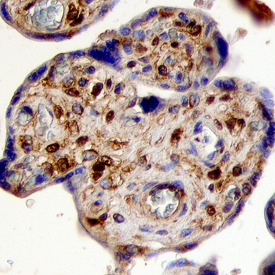
- FGF-5 in Human Placenta. FGF-5 was detected in immersion fixed paraffin-embedded sections of human placenta using Goat Anti-Human FGF-5 Antigen Affinity-purified Polyclonal Antibody (Catalog # AF-237-NA) at 10 µg/mL overnight at 4 °C. Before incubation with the primary antibody, tissue was subjected to heat-induced epitope retrieval using Antigen Retrieval Reagent-Basic (Catalog # CTS013). Tissue was stained using the Anti-Goat HRP-DAB Cell & Tissue Staining Kit (brown; Catalog # CTS008) and counterstained with hematoxylin (blue). Specific staining was localized to trophoblast cells in chorionic villi. View our protocol for Chromogenic IHC Staining of Paraffin-embedded Tissue Sections.
Supportive validation
- Submitted by
- R&D Systems (provider)
- Main image
- Experimental details
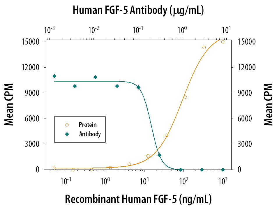
- Cell Proliferation Induced by FGF-5 and Neutralization by Human FGF-5 Antibody. Recombinant Human FGF-5 (Catalog # 237-F5) stimulates proliferation in the the NR6R-3T3 mouse fibroblast cell line in a dose-dependent manner (orange line). Proliferation elicited by Recombinant Human FGF-5 (20 ng/mL) is neutralized (green line) by increasing concentrations of Goat Anti-Human FGF-5 Antigen Affinity-purified Polyclonal Antibody (Catalog # AF-237-NA). The ND50 is typically 0.2-0.8 µg/mL in the presence of heparin (1 µg/mL).