 Explore
 Validate
 Learn
Learn Western blot
Antibody data
- Antibody Data
- Antigen structure
- References [1]
- Comments [0]
- Validations
- Western blot [1]
- Immunohistochemistry [1]
Submit
Validation data
Reference
Comment
Report error
- Product number
- AF6224 - Provider product page

- Provider
- R&D Systems
- Product name
- Mouse FGF-10 Antibody
- Antibody type
- Polyclonal
- Description
- Antigen Affinity-purified. Detects mouse and human FGF-10 in Western blots. In direct ELISAs, approximately 100% cross-reactivity with recombinant rat FGF-10 is observed and less than 10% cross-reactivity with recombinant mouse FGF-6 and recombinant mouse FGF-7 is observed.
- Reactivity
- Mouse
- Host
- Sheep
- Conjugate
- Unconjugated
- Antigen sequence
NP_032028- Isotype
- IgG
- Vial size
- 100 ug
- Concentration
- LYOPH
- Storage
- Use a manual defrost freezer and avoid repeated freeze-thaw cycles. 12 months from date of receipt, -20 to -70 °C as supplied. 1 month, 2 to 8 °C under sterile conditions after reconstitution. 6 months, -20 to -70 °C under sterile conditions after reconstitution.
Submitted references Generation of iPSC-derived limb progenitor-like cells for stimulating phalange regeneration in the adult mouse.
Chen Y, Xu H, Lin G
Cell discovery 2017;3:17046
Cell discovery 2017;3:17046
No comments: Submit comment
Supportive validation
- Submitted by
- R&D Systems (provider)
- Main image
- Experimental details
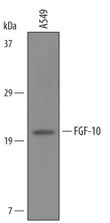
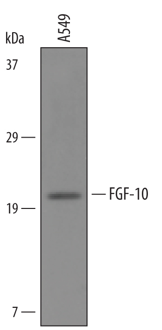
- Detection of Human FGF-10 by Western Blot. Western blot shows lysates of A549 human lung carcinoma cell line. PVDF Membrane was probed with 1 µg/mL of Sheep Anti-Mouse FGF-10 Antigen Affinity-purified Polyclonal Antibody (Catalog # AF6224) followed by HRP-conjugated Anti-Sheep IgG Secondary Antibody (Catalog # HAF016). A specific band was detected for FGF-10 at approximately 20 kDa (as indicated). This experiment was conducted under reducing conditions and using Immunoblot Buffer Group 8.
Supportive validation
- Submitted by
- R&D Systems (provider)
- Main image
- Experimental details
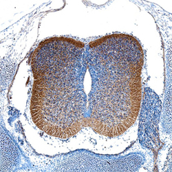
- FGF-10 in Mouse Embryo. FGF-10 was detected in immersion fixed frozen sections of mouse embryo (E13) using Sheep Anti-Mouse FGF-10 Antigen Affinity-purified Polyclonal Antibody (Catalog # AF6224) at 5 µg/mL overnight at 4 °C. Tissue was stained using the Anti-Sheep HRP-DAB Cell & Tissue Staining Kit (brown; Catalog # CTS019) and counterstained with hematoxylin (blue). Specific staining was localized to developing spinal cord. View our protocol for Chromogenic IHC Staining of Frozen Tissue Sections.
