Explore
 Validate
 Learn
Learn Western blot
 Immunoprecipitation
Antibody data
- Antibody Data
- Antigen structure
- References [0]
- Comments [0]
- Validations
- Western blot [2]
- Immunoprecipitation [3]
- Immunohistochemistry [1]
Submit
Validation data
Reference
Comment
Report error
- Product number
- LS-C99724 - Provider product page

- Provider
- LSBio
- Proper citation
- LifeSpan Cat#LS-C99724, RRID:AB_2163922
- Product name
- PIM2 / Pim-2 Antibody (aa277-308) LS-C99724
- Antibody type
- Polyclonal
- Description
- Ammonium sulfate precipitation
- Reactivity
- Human
- Host
- Rabbit
- Storage
- Maintain refrigerated at 2°C to 8°C for up to 6 months. For long term storage store at -20°C.
No comments: Submit comment
Enhanced validation
- Submitted by
- LSBio (provider)
- Enhanced method
- Genetic validation
- Main image

- Experimental details
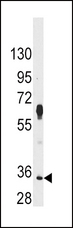
- Western blot of anti-PIM2 Antibody in HeLa cell line lysates (35 ug/lane). PIM2 (arrow) was detected using the purified antibody.
- Submitted by
- LSBio (provider)
- Enhanced method
- Genetic validation
- Main image

- Experimental details
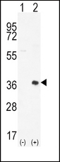
- Western blot of PIM2 (arrow) using rabbit polyclonal PIM2 Antibody (D292). 293 cell lysates (2 ug/lane) either nontransfected (Lane 1) or transiently transfected (Lane 2) with the PIM2 gene.
Supportive validation
- Submitted by
- LSBio (provider)
- Enhanced method
- Genetic validation
- Main image

- Experimental details
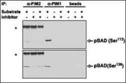
- PIM proteins were immunoprecipitated from MV4;11 cells and the agarose-protein A-immunoprecipitate complex was tested for its ability to phosphorylate BAD in vitro in the presence or absence of K00135. Phosphorylation of BAD (both on Ser112 and Ser136, detected by WB with phospho-specific antibodies) was abrogated on addition of the compound. Asterisks, strong bands corresponding to the heavy chain of the anti-PIM2 rabbit antibody recognized by the antirabbit immunoglobulin G secondary antibody. Beads alone (without anti-PIM antibodies) were incubated with the MV4;11 extract and used for the same in vitro phosphorylation reaction as a negative control.

- Submitted by
- LSBio (provider)
- Main image
- Experimental details
- PIM proteins were immunoprecipitated from MV4;11 cells and the agarose-protein A-immunoprecipitate complex was tested for its ability to phosphorylate BAD in vitro in the presence or absence of K00135. Phosphorylation of BAD (both on Ser112 and Ser136, detected by WB with phospho-specific antibodies) was abrogated on addition of the compound. Asterisks, strong bands corresponding to the heavy chain of the anti-PIM2 rabbit antibody recognized by the antirabbit immunoglobulin G secondary antibody. Beads alone (without anti-PIM antibodies) were incubated with the MV4;11 extract and used for the same in vitro phosphorylation reaction as a negative control.

- Submitted by
- LSBio (provider)
- Main image

- Experimental details
- PIM proteins were immunoprecipitated from MV4;11 cells and the agarose-protein A-immunoprecipitate complex was tested for its ability to phosphorylate BAD in vitro in the presence or absence of K00135. Phosphorylation of BAD (both on Ser112 and Ser136, detected by WB with phospho-specific antibodies) was abrogated on addition of the compound. Asterisks, strong bands corresponding to the heavy chain of the anti-PIM2 rabbit antibody recognized by the antirabbit immunoglobulin G secondary antibody. Beads alone (without anti-PIM antibodies) were incubated with the MV4;11 extract and used for the same in vitro phosphorylation reaction as a negative control.

Supportive validation
- Submitted by
- LSBio (provider)
- Enhanced method
- Genetic validation
- Main image
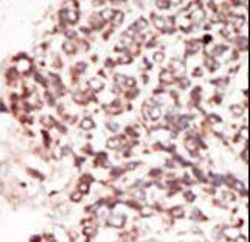
- Experimental details
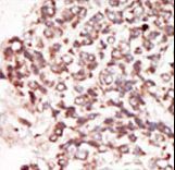
- Formalin-fixed and paraffin-embedded human cancer tissue reacted with the primary antibody, which was peroxidase-conjugated to the secondary antibody, followed by DAB staining. This data demonstrates the use of this antibody for immunohistochemistry; clinical relevance has not been evaluated. BC = breast carcinoma; HC = hepatocarcinoma.