Explore
 Validate
 Learn
Learn Western blot
Antibody data
- Antibody Data
- Antigen structure
- References [1]
- Comments [0]
- Validations
- Western blot [2]
- Immunocytochemistry [1]
- Immunoprecipitation [1]
- Immunohistochemistry [3]
- Flow cytometry [1]
Submit
Validation data
Reference
Comment
Report error
- Product number
- GTX83892 - Provider product page

- Provider
- GeneTex
- Proper citation
- GeneTex Cat#GTX83892, RRID:AB_10726848
- Product name
- PIM2 antibody [9A5]
- Antibody type
- Monoclonal
- Reactivity
- Human
- Host
- Mouse
Submitted references PIM2-mediated phosphorylation of hexokinase 2 is critical for tumor growth and paclitaxel resistance in breast cancer.
Yang T, Ren C, Qiao P, Han X, Wang L, Lv S, Sun Y, Liu Z, Du Y, Yu Z
Oncogene 2018 Nov;37(45):5997-6009
Oncogene 2018 Nov;37(45):5997-6009
No comments: Submit comment
Supportive validation
- Submitted by
- GeneTex (provider)
- Main image
- Experimental details
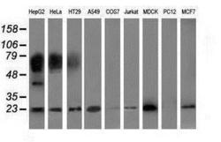
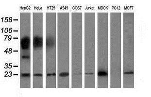
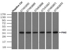
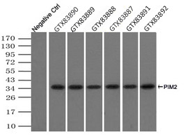
- Western blot analysis of extracts (35ug) from 9 different cell lines by using anti-PIM2 monoclonal antibody.
- Submitted by
- GeneTex (provider)
- Main image
- Experimental details
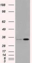
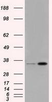
- HEK293T cells were transfected with the pCMV6-ENTRY control (Left lane) or pCMV6-ENTRY PIM2 (Right lane) cDNA for 48 hrs and lysed. Equivalent amounts of cell lysates (5 ug per lane) were separated by SDS-PAGE and immunoblotted with anti-PIM2.
Supportive validation
- Submitted by
- GeneTex (provider)
- Main image
- Experimental details
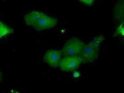
- Anti-PIM2 mouse monoclonal antibody (GTX83892) immunofluorescent staining of COS7 cells transiently transfected with PIM2

Supportive validation
- Submitted by
- GeneTex (provider)
- Main image
- Experimental details
- Immunoprecipitation(IP) of PIM2 by using TrueMab monoclonal anti-PIM2 antibodies (Negative control: IP without adding anti-PIM2 antibody). For each experiment, 500ul of DDK tagged PIM2 overexpression lysates (at 1:5 dilution with HEK293T lysate), 2ug of anti-PIM2 antibody and 20ul (0.1mg) of goat anti-mouse conjugated magnetic beads were mixed and incubated overnight. After extensive wash to remove any non-specific binding, the immuno-precipitated products were analyzed with rabbit anti-DDK polyclonal antibody.
Supportive validation
- Submitted by
- GeneTex (provider)
- Main image

- Experimental details
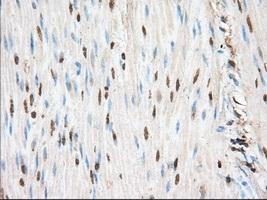
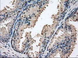
- Immunohistochemical staining of paraffin-embedded Human colon tissue using anti-PIM2 mouse monoclonal antibody. (GTX83892, Dilution 1:50)
- Submitted by
- GeneTex (provider)
- Main image
- Experimental details
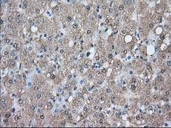
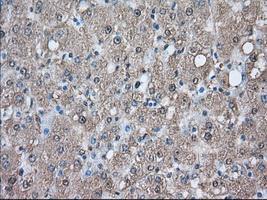
- Immunohistochemical staining of paraffin-embedded Human liver tissue using anti-PIM2 mouse monoclonal antibody. (GTX83892, Dilution 1:50)
- Submitted by
- GeneTex (provider)
- Main image
- Experimental details
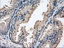
- Immunohistochemical staining of paraffin-embedded Human prostate tissue using anti-PIM2 mouse monoclonal antibody. (GTX83892, Dilution 1:50)
Supportive validation
- Submitted by
- GeneTex (provider)
- Main image
- Experimental details
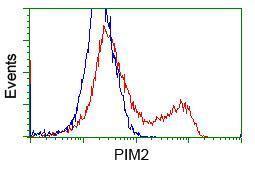
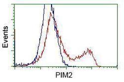
- HEK293T cells transfected with either RC201933 overexpress plasmid(Red) or empty vector control plasmid(Blue) were immunostained by anti-PIM2 antibody(GTX83892), and then analyzed by flow cytometry.