Explore
 Validate
 Learn
Learn Immunohistochemistry
Antibody data
- Antibody Data
- Antigen structure
- References [4]
- Comments [0]
- Validations
- Immunohistochemistry [1]
Submit
Validation data
Reference
Comment
Report error
- Product number
- HPA021555 - Provider product page

- Provider
- Atlas Antibodies
- Proper citation
- Atlas Antibodies Cat#HPA021555, RRID:AB_1851969
- Product name
- Anti-JAG1
- Antibody type
- Polyclonal
- Description
- Polyclonal Antibody against Human JAG1, Gene description: jagged 1, Alternative Gene Names: AGS, AHD, AWS, CD339, HJ1, JAGL1, Validated applications: IHC, Uniprot ID: P78504, Storage: Store at +4°C for short term storage. Long time storage is recommended at -20°C.
- Reactivity
- Human
- Host
- Rabbit
- Conjugate
- Unconjugated
- Isotype
- IgG
- Vial size
- 100 µl
- Concentration
- 0.3 mg/ml
- Storage
- Store at +4°C for short term storage. Long time storage is recommended at -20°C.
- Handling
- The antibody solution should be gently mixed before use.
Submitted references IL-4-dependent Jagged1 expression/processing is associated with survival of chronic lymphocytic leukemia cells but not with Notch activation
Knockdown of L1CAM significantly reduces metastasis in a xenograft model of human melanoma: L1CAM is a potential target for anti-melanoma therapy
Endothelial follistatin‐like‐1 regulates the postnatal development of the pulmonary vasculature by modulating BMP/Smad signaling
Increasing diagnostic accuracy to grade dysplasia in Barrett’s esophagus using an immunohistochemical panel for CDX2, p120ctn, c-Myc and Jagged1
De Falco F, Del Papa B, Baldoni S, Sabatini R, Falzetti F, Di Ianni M, Martelli M, Mezzasoma F, Pelullo M, Marconi P, Sportoletti P, Screpanti I, Rosati E
Cell Death & Disease 2018;9(12)
Cell Death & Disease 2018;9(12)
Knockdown of L1CAM significantly reduces metastasis in a xenograft model of human melanoma: L1CAM is a potential target for anti-melanoma therapy
Mattei F, Ernst A, Putscher A, Samatov T, Suling A, Galatenko V, Shkurnikov M, Knyazev E, Tonevitsky A, Haalck T, Lange T, Maar H, Schröder- Schwarz J, Riecken K, Schumacher U, Wicklein D
PLOS ONE 2018;13(2):e0192525
PLOS ONE 2018;13(2):e0192525
Endothelial follistatin‐like‐1 regulates the postnatal development of the pulmonary vasculature by modulating BMP/Smad signaling
Tania N, Maarsingh H, T. Bos I, Mattiotti A, Prakash S, Timens W, Gunst Q, Jimenez‐Borreguero L, Schmidt M, van den Hoff M, Gosens R
Pulmonary Circulation 2017;7(1):219-231
Pulmonary Circulation 2017;7(1):219-231
Increasing diagnostic accuracy to grade dysplasia in Barrett’s esophagus using an immunohistochemical panel for CDX2, p120ctn, c-Myc and Jagged1
Karamchandani D, Lehman H, Ohanessian S, Massé J, Welsh P, Odze R, Goldblum J, Berg A, Stairs D
Diagnostic Pathology 2016;11(1)
Diagnostic Pathology 2016;11(1)
No comments: Submit comment
Supportive validation
- Submitted by
- Atlas Antibodies (provider)
- Enhanced method
- Orthogonal validation
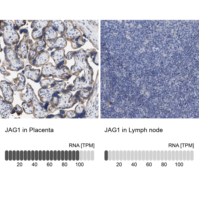
- Main image
- Experimental details
- Immunohistochemistry analysis in human placenta and lymph node tissues using HPA021555 antibody. Corresponding JAG1 RNA-seq data are presented for the same tissues.
- Sample type
- Human
- Protocol
- Protocol