 Explore
 Validate
 Learn
Learn Western blot
 ELISA
ELISAAntibody data
- Antibody Data
- Antigen structure
- References [0]
- Comments [0]
- Validations
- Western blot [1]
- Immunocytochemistry [1]
- Immunohistochemistry [1]
Submit
Validation data
Reference
Comment
Report error
- Product number
- AP09127PU-N - Provider product page

- Provider
- Acris Antibodies GmbH
- Proper citation
- Acris Antibodies GmbH Cat#AP09127PU-N, RRID:AB_2035312
- Product name
- anti CD339 / JAG1 (110-125)
- Antibody type
- Polyclonal
- Antigen
- Synthetic peptide corresponding to amino acids 110-125 of Human Jagged-1 protein.
- Reactivity
- Human, Mouse, Rat
- Host
- Rabbit
- Isotype
- IgG
- Vial size
- 0.5 mg
- Concentration
- 1.0 mg/ml (by UV absorbance at 280 nm)
No comments: Submit comment
Supportive validation
- Submitted by
- Acris Antibodies GmbH (provider)
- Main image
- Experimental details
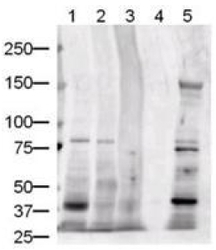
- Western blot using AP09127PU-N Jagged-1 antibody at 1/500 dilution shows detection of Jagged-1 protein in various whole cell lysates (20 µg/Lane). The membrane was washed and reacted with a 1/5,000 dilution of HRP conjugated Goat anti-Rabbit IgG. The band at ~134 kDa in Lane 5 is believed to be Jagged-1 precursor. The identity of minor reactive bands is unknown.Lane 1: Human brain. Lane 2: Human kidney. Lane 3: Human liver. Lane 4: Sample buffer only.Lane 5: Mouse liver. Exposure time was 1 min.Predicted MW: 134 kDa.

Supportive validation
- Submitted by
- Acris Antibodies GmbH (provider)
- Main image

- Experimental details
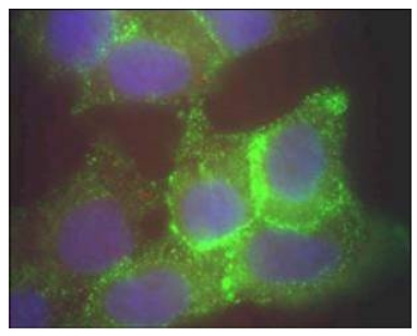
- Immunofluorescence Microscopy using AP09127PU-N Jagged-1 antibody of Human corneal epithelial cells. Primary antibody was used at a 1/500 dilution. The Jagged-1 (green staining) is localized to the cytoplasm and is consistent with reports in the literature. The nucleus is stained with Bis benzimide (blue). Personal Communication. Aihua Ma, Univdersity of Cardiff.
Supportive validation
- Submitted by
- Acris Antibodies GmbH (provider)
- Main image
- Experimental details
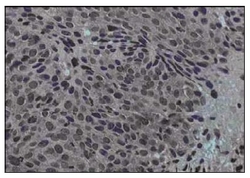
- AP09127PU-N Jagged-1 antibody Immunohistochemical staining of Formalin-Fixed, Paraffin Embedded Human cervical cancer tissue (40X magnification). Hematoxylin was used to counter-stain cells. A 1/100 dilution of primary antibody was used. Personal Communication. Martin Kast Laboratory.
