Explore
 Validate
 Learn
Learn Western blot
Antibody data
- Antibody Data
- Antigen structure
- References [0]
- Comments [0]
- Validations
- Western blot [3]
- Immunohistochemistry [2]
Submit
Validation data
Reference
Comment
Report error
- Product number
- PA5-27875 - Provider product page

- Provider
- Invitrogen Antibodies
- Product name
- Anti-FRS2 Polyclonal Antibody
- Antibody type
- Polyclonal
- Antigen
- Recombinant protein fragment
- Description
- Recommended positive controls: PC-3. Predicted reactivity: Mouse (97%), Rat (96%), Xenopus laevis (87%), Pig (99%), Chicken (97%). Store product as a concentrated solution. Centrifuge briefly prior to opening the vial.
- Reactivity
- Human, Mouse
- Host
- Rabbit
- Isotype
- IgG
- Vial size
- 100 µL
- Concentration
- 1 mg/mL
- Storage
- Store at 4°C short term. For long term storage, store at -20°C, avoiding freeze/thaw cycles.
No comments: Submit comment
Supportive validation
- Submitted by
- Invitrogen Antibodies (provider)
- Main image

- Experimental details
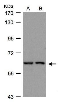
- Western blot analysis of FRS2 using 30 µg of A) H1299 and B) HeLa S3 lysate. Samples were loaded onto a 7.5% SDS-PAGE gel and probed with a FRS2 polyclonal antibody (Product # PA5-27875) at a dilution of 1:500.
- Submitted by
- Invitrogen Antibodies (provider)
- Main image
- Experimental details
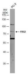
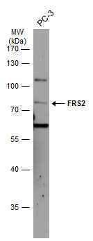
- Western blot analysis of FRS2 was performed by separating 30 µg of whole cell extract by 10% SDS-PAGE. Proteins were transferred to a membrane and probed with a FRS2 Polyclonal Antibody (Product # PA5-27875) at a dilution of 1:500. The HRP-conjugated anti-rabbit IgG antibody was used to detect the primary antibody.
- Submitted by
- Invitrogen Antibodies (provider)
- Main image
- Experimental details
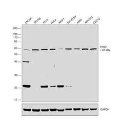
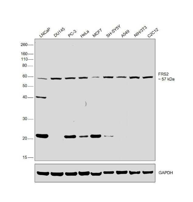
- Western blot was performed using Anti-FRS2 Polyclonal Antibody (Product # PA5-27875) and a 57kDa band corresponding to FRS2 was observed across cell lines tested. Whole cell extracts (30 µg lysate) of LNCaP (Lane 1), DU145 (Lane 2), PC-3 (Lane 3), HeLa (Lane 4), MCF7 (Lane 5), SH-SY5Y (Lane 6), A549 (Lane 7), NIH/3T3 (Lane 8) and C2C12 (Lane 9) were electrophoresed using NuPAGE™ 4-12% Bis-Tris Protein Gel (Product # NP0322BOX). Resolved proteins were then transferred onto a nitrocellulose membrane (Product # IB23001) by iBlot® 2 Dry Blotting System (Product # IB21001). The blot was probed with the primary antibody (1:500 dilution) and detected by chemiluminescence Goat anti-Rabbit IgG (H+L) Superclonal™ Secondary Antibody, HRP (Product # A27036, 1:4000 dilution) using the iBright FL 1000 (Product # A32752). Chemiluminescent detection was performed using Novex® ECL Chemiluminescent Substrate Reagent Kit (Product # WP20005).
Supportive validation
- Submitted by
- Invitrogen Antibodies (provider)
- Main image

- Experimental details
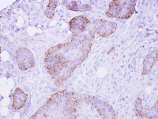
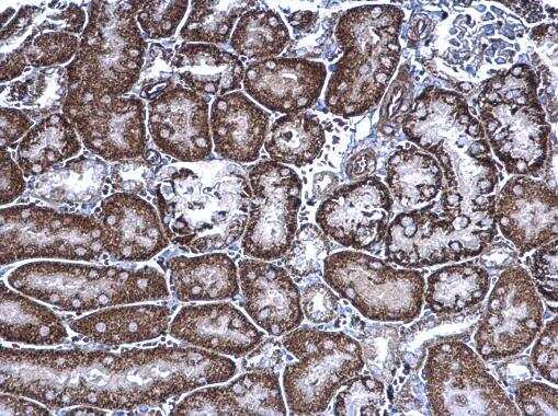
- FRS2 Polyclonal Antibody detects FRS2 protein at cytoplasm on mouse kidney by immunohistochemical analysis. Sample: Paraffin-embedded mouse kidney. FRS2 Polyclonal Antibody (Product # PA5-27875) diluted at 1:500. Antigen Retrieval: EDTA based buffer, pH 8.0, 15 min.
- Submitted by
- Invitrogen Antibodies (provider)
- Main image

- Experimental details
- FRS2 Polyclonal Antibody detects FRS2 protein at membrane on HSC-3 xenograft by immunohistochemical analysis. Sample: Paraffin-embedded HSC-3 xenograft. FRS2 Polyclonal Antibody (Product # PA5-27875) dilution: 1:500. Antigen Retrieval: EDTA based buffer, pH 8.0, 15 min.