 Explore
 Validate
 Learn
Learn Western blot
 Immunohistochemistry
Antibody data
- Antibody Data
- Antigen structure
- References [7]
- Comments [0]
- Validations
- Immunohistochemistry [3]
- Other assay [4]
Submit
Validation data
Reference
Comment
Report error
- Product number
- 38-5200 - Provider product page

- Provider
- Invitrogen Antibodies
- Product name
- SOCS1 Polyclonal Antibody
- Antibody type
- Polyclonal
- Antigen
- Other
- Reactivity
- Human, Mouse
- Host
- Rabbit
- Isotype
- IgG
- Vial size
- 100 μg
- Concentration
- 0.25 mg/mL
- Storage
- -20°C
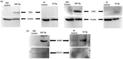
Submitted references Implication of Toll/IL-1 receptor domain containing adapters in Porphyromonas gingivalis-induced inflammation.
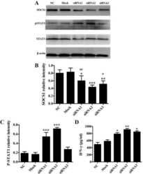
N(6)-Adenosine Methylation of Socs1 mRNA Is Required to Sustain the Negative Feedback Control of Macrophage Activation.
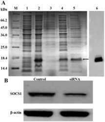
Silencing of suppressor of cytokine signaling 1 enhances the immunological effect of mucin 1-calreticulin-primed 4T1 cell-treated dendritic cells in breast cancer treatment.
Induction of miR-155 after Brain Injury Promotes Type 1 Interferon and has a Neuroprotective Effect.
Signal Transducer and Activator of Transcription 1 (STAT1) Knock-down Induces Apoptosis in Malignant Pleural Mesothelioma.
1,25-Dihydroxyvitamin D promotes negative feedback regulation of TLR signaling via targeting microRNA-155-SOCS1 in macrophages.
Stage dependent aberrant regulation of cytokine-STAT signaling in murine systemic lupus erythematosus.
Bugueno IM, Benkirane-Jessel N, Huck O
Innate immunity 2021 May;27(4):324-342
Innate immunity 2021 May;27(4):324-342
N(6)-Adenosine Methylation of Socs1 mRNA Is Required to Sustain the Negative Feedback Control of Macrophage Activation.
Du J, Liao W, Liu W, Deb DK, He L, Hsu PJ, Nguyen T, Zhang L, Bissonnette M, He C, Li YC
Developmental cell 2020 Dec 21;55(6):737-753.e7
Developmental cell 2020 Dec 21;55(6):737-753.e7
Silencing of suppressor of cytokine signaling 1 enhances the immunological effect of mucin 1-calreticulin-primed 4T1 cell-treated dendritic cells in breast cancer treatment.
Qin S, Gao Z, Liu Y, Liu C, Wang J, Zou LL
Oncology letters 2018 Feb;15(2):1630-1638
Oncology letters 2018 Feb;15(2):1630-1638
Induction of miR-155 after Brain Injury Promotes Type 1 Interferon and has a Neuroprotective Effect.
Harrison EB, Emanuel K, Lamberty BG, Morsey BM, Li M, Kelso ML, Yelamanchili SV, Fox HS
Frontiers in molecular neuroscience 2017;10:228
Frontiers in molecular neuroscience 2017;10:228
Signal Transducer and Activator of Transcription 1 (STAT1) Knock-down Induces Apoptosis in Malignant Pleural Mesothelioma.
Arzt L, Halbwedl I, Gogg-Kamerer M, Popper HH
Pathology oncology research : POR 2017 Jul;23(3):595-605
Pathology oncology research : POR 2017 Jul;23(3):595-605
1,25-Dihydroxyvitamin D promotes negative feedback regulation of TLR signaling via targeting microRNA-155-SOCS1 in macrophages.
Chen Y, Liu W, Sun T, Huang Y, Wang Y, Deb DK, Yoon D, Kong J, Thadhani R, Li YC
Journal of immunology (Baltimore, Md. : 1950) 2013 Apr 1;190(7):3687-95
Journal of immunology (Baltimore, Md. : 1950) 2013 Apr 1;190(7):3687-95
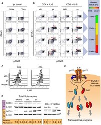
Stage dependent aberrant regulation of cytokine-STAT signaling in murine systemic lupus erythematosus.
Hale MB, Krutzik PO, Samra SS, Crane JM, Nolan GP
PloS one 2009 Aug 25;4(8):e6756
PloS one 2009 Aug 25;4(8):e6756
No comments: Submit comment
Supportive validation
- Submitted by
- Invitrogen Antibodies (provider)
- Main image
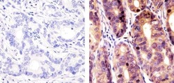
- Experimental details
- Immunohistochemistry analysis of SOCS-1 showing staining in the cytoplasm and nucleus of paraffin-embedded human stomach carcinoma (right) compared to a negative control without primary antibody (left). To expose target proteins, antigen retrieval was performed using 10mM sodium citrate (pH 6.0), microwaved for 8-15 min. Following antigen retrieval, tissues were blocked in 3% H2O2-methanol for 15 min at room temperature, washed with ddH2O and PBS, and then probed with a Anti- SOCS-1 Polyclonal Antibody (Product # 38-5200) diluted in 3% BSA-PBS at a dilution of 1:20 overnight at 4°C in a humidified chamber. Tissues were washed extensively in PBST and detection was performed using an HRP-conjugated secondary antibody followed by colorimetric detection using a DAB kit. Tissues were counterstained with hematoxylin and dehydrated with ethanol and xylene to prep for mounting.

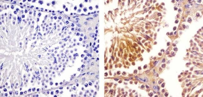
- Submitted by
- Invitrogen Antibodies (provider)
- Main image
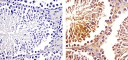
- Experimental details
- Immunohistochemistry analysis of SOCS-1 showing staining in the cytoplasm and nucleus of paraffin-embedded mouse testis tissue (right) compared to a negative control without primary antibody (left). To expose target proteins, antigen retrieval was performed using 10mM sodium citrate (pH 6.0), microwaved for 8-15 min. Following antigen retrieval, tissues were blocked in 3% H2O2-methanol for 15 min at room temperature, washed with ddH2O and PBS, and then probed with a Anti- SOCS-1 Polyclonal Antibody (Product # 38-5200) diluted in 3% BSA-PBS at a dilution of 1:20 overnight at 4°C in a humidified chamber. Tissues were washed extensively in PBST and detection was performed using an HRP-conjugated secondary antibody followed by colorimetric detection using a DAB kit. Tissues were counterstained with hematoxylin and dehydrated with ethanol and xylene to prep for mounting.
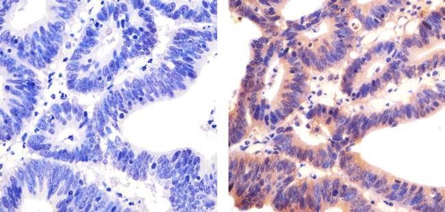
- Submitted by
- Invitrogen Antibodies (provider)
- Main image

- Experimental details
- Immunohistochemistry analysis of SOCS-1 showing staining in the cytoplasm and nucleus of paraffin-embedded human colon carcinoma (right) compared to a negative control without primary antibody (left). To expose target proteins, antigen retrieval was performed using 10mM sodium citrate (pH 6.0), microwaved for 8-15 min. Following antigen retrieval, tissues were blocked in 3% H2O2-methanol for 15 min at room temperature, washed with ddH2O and PBS, and then probed with a Anti- SOCS-1 Polyclonal Antibody (Product # 38-5200) diluted in 3% BSA-PBS at a dilution of 1:20 overnight at 4°C in a humidified chamber. Tissues were washed extensively in PBST and detection was performed using an HRP-conjugated secondary antibody followed by colorimetric detection using a DAB kit. Tissues were counterstained with hematoxylin and dehydrated with ethanol and xylene to prep for mounting.
Supportive validation
- Submitted by
- Invitrogen Antibodies (provider)
- Main image
- Experimental details
- NULL

- Submitted by
- Invitrogen Antibodies (provider)
- Main image
- Experimental details
- NULL

- Submitted by
- Invitrogen Antibodies (provider)
- Main image
- Experimental details
- Figure 4 Differential inhibition of pStat1 and pStat3 signaling responses to IL-6 correlates with increased expression of SOCS1 protein. (A) Basal phosphorylation of Stat1 and Stat3 in B220-, CD4 and CD8 single positive T cells from lpr mice at 5, 10, 15, and 20 weeks of age. The same subsets from MRL mice have basal signaling that does not change with age and resembles that of 5-week-old lpr mice (not shown). (B) Phosphorylation of Stat1 and Stat3 in CD4 and CD8 single positive T cells after stimulation with IL-6. Nearly all single positive T cells from MRL mice show strong phosphorylation of Stat1 and Stat3 after stimulation with IL-6 and that did not change with age. In lpr mice, starting at 10 weeks, a large proportion of CD8+ T cells lost the pStat1 response to IL-6 but maintained pStat3 response to this cytokine at every age. Cells are in the upper left quadrant because, at the single-cell level, there is strong IL-6-induced phospho-activation of Stat3, but little or no detectable phospho-activation of Stat1. CD4+ T cells from 10-week-old lpr mice had no detectable pStat1 response to IL-6 and at later time points an increasing proportion of CD4+ T cells lost the pStat3 response as well. To the right of these two-dimensional plots is a set of color bars that summarize the severity of SLE in the lpr mice through time. (C) Flow cytometric analysis of IL-6 receptor expression on CD4 and CD8 single positive T cells at 20 weeks of age. (D) Western blot analysis of SOCS1 prote

- Submitted by
- Invitrogen Antibodies (provider)
- Main image
- Experimental details
- Figure 4. P. gingivalis induced the activation of TLR2 and TLR4, activation of the SARM adapter, and the inhibition of SOCS-1 protein expression. (A) Protein expression of TLR2, TLR4 in response to P. gingivalis ( Pg ) (MOI:100) for 24 h was evaluated by Western blot. (B) Protein expression of SARM and SOCS-1 in response to P. gingivalis (MOI:100) for 24 h in GEC and EC was evaluated by Western blot. All bands were compared and normalized with beta-actin expression.
