Explore
 Validate
 Learn
Learn Western blot
Antibody data
- Antibody Data
- Antigen structure
- References [1]
- Comments [0]
- Validations
- Western blot [1]
- Immunohistochemistry [1]
- Other assay [1]
Submit
Validation data
Reference
Comment
Report error
- Product number
- PA5-28533 - Provider product page

- Provider
- Invitrogen Antibodies
- Product name
- PHF16 Polyclonal Antibody
- Antibody type
- Polyclonal
- Antigen
- Recombinant full-length protein
- Description
- Recommended positive controls: 293T, HepG2. Store product as a concentrated solution. Centrifuge briefly prior to opening the vial.
- Reactivity
- Human
- Host
- Rabbit
- Isotype
- IgG
- Vial size
- 100 μL
- Concentration
- 0.62 mg/mL
- Storage
- Store at 4°C short term. For long term storage, store at -20°C, avoiding freeze/thaw cycles.
Submitted references Dynamics of the Transcriptome during Human Spermatogenesis: Predicting the Potential Key Genes Regulating Male Gametes Generation.
Zhu Z, Li C, Yang S, Tian R, Wang J, Yuan Q, Dong H, He Z, Wang S, Li Z
Scientific reports 2016 Jan 12;6:19069
Scientific reports 2016 Jan 12;6:19069
No comments: Submit comment
Supportive validation
- Submitted by
- Invitrogen Antibodies (provider)
- Main image

- Experimental details
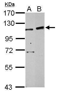
- Western Blot using PHF16 Polyclonal Antibody (Product # PA5-28533). Sample (30 µg of whole cell lysate). Lane A: 293T. Lane B: HepG2 . 7.5% SDS PAGE. PHF16 Polyclonal Antibody (Product # PA5-28533) diluted at 1:500.
Supportive validation
- Submitted by
- Invitrogen Antibodies (provider)
- Main image

- Experimental details
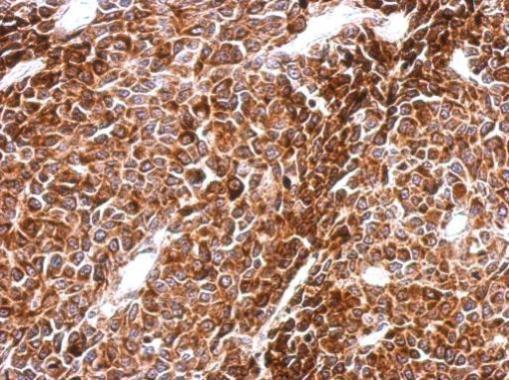
- Immunohistochemical analysis of paraffin-embedded BT483 xenograft, using PHF16 (Product # PA5-28533) antibody at 1:500 dilution. Antigen Retrieval: EDTA based buffer, pH 8.0, 15 min.
Supportive validation
- Submitted by
- Invitrogen Antibodies (provider)
- Main image

- Experimental details
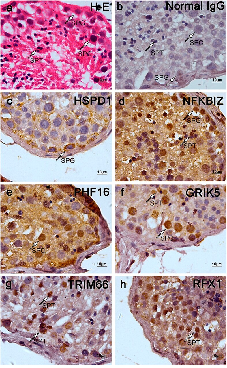
- Figure 4 Immunohistochemistry certified the localization of the proteins of corresponding genes in testicular tissues. A total of 6 genes were selected to be detected on testicular tissue sections derived from OA patients with normal spermatogenesis via immunohistochemistry, normal Rabbit IgG was used as primary antibody for negative control. Typical germ cells were pointed by arrows. ( a ) Hematoxylin-eosin (H-E) staining of testicular tissue section. Normal spermatogenesis could be observed; ( b ) Negative control stained with normal IgG. Though there was a little non-specific background staining, no specific staining could be observed; ( c ) Section stained with anti-HSP60 (HSPD1) antibody. HSPD1 belongs to Cluster 1. (See Figs 2d and 3a ) Spermatogonial Cells were stained in brown; ( d ) Section stained with anti-NFKBIZ antibody. NFKBIZ belongs to Cluster 2. Spermatogonial cells and some spermatids were stained in brown; ( e ) Section stained with anti-PHF16 (JADE3) antibody, PHF16 belongs to Cluster3. Spermatocytes were stained in brown; ( f ) Section stained with anti-GRIK5 antibody. GRIK5 belongs to Cluster 3. Many germ cells were stained in brown, among which spermatocytes and some spermatids were stained in a relatively dark color; ( g ) Section stained with anti-TRIM66 antibody. TRIM66 belongs to Cluster 4. Spermatids were stained in brown; ( h ) Section stained with anti-RFX1 antibody. RFX1 belongs to Cluster 4. Spermatids were stained in brown. These distribution