Explore
 Validate
 Learn
LearnPA5-20268
antibody from Invitrogen Antibodies
Targeting: BCL2A1
ACC-1, ACC-2, ACC1, ACC2, BCL2L5, BFL1, GRS, HBPA1
 Western blot
Antibody data
- Antibody Data
- Antigen structure
- References [1]
- Comments [0]
- Validations
- Western blot [1]
- Immunocytochemistry [1]
- Immunohistochemistry [3]
- Other assay [2]
Submit
Validation data
Reference
Comment
Report error
- Product number
- PA5-20268 - Provider product page

- Provider
- Invitrogen Antibodies
- Product name
- BCL2A1 Polyclonal Antibody
- Antibody type
- Polyclonal
- Antigen
- Synthetic peptide
- Description
- Despite the predicted molecular weight, Bfl-1 often migrates at a higher molecular weight in SDS-PAGE, presumably due to post-translational modifications. A suggested positive control is human kidney tissue lysate. PA5-20268 can be used with blocking peptide PEP-0383.
- Reactivity
- Human, Mouse
- Host
- Rabbit
- Isotype
- IgG
- Vial size
- 100 μg
- Concentration
- 1 mg/mL
- Storage
- Maintain refrigerated at 2-8°C for up to 3 months. For long term storage store at -20°C
Submitted references Yaobishu Regulates Inflammatory, Metabolic, Autophagic, and Apoptosis Pathways to Attenuate Lumbar Disc Herniation.
Li X, Li S, Zang Z, He Y
Oxidative medicine and cellular longevity 2022;2022:3861380
Oxidative medicine and cellular longevity 2022;2022:3861380
No comments: Submit comment
Supportive validation
- Submitted by
- Invitrogen Antibodies (provider)
- Main image
- Experimental details
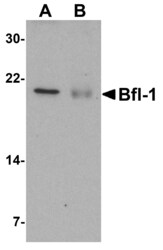
- Western Blot analysis of Bfl-1 in (A) human kidney and (B) human lung tissue lysate with BCL2A1 Polyclonal Antibody (Product # PA5-20268) at 1 µg/mL.

Supportive validation
- Submitted by
- Invitrogen Antibodies (provider)
- Main image
- Experimental details
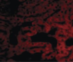
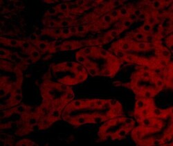
- Immunofluorescent analysis of mouse kidney cells using a Bfl-1 polyclonal antibody (Product # PA5-20268) at a 20 µg/mL dilution.
Supportive validation
- Submitted by
- Invitrogen Antibodies (provider)
- Main image
- Experimental details
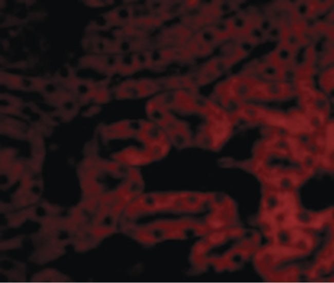
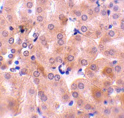
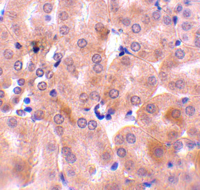
- Immunohistochemistry of Bfl-1 in mouse kidney tissue with BCL2A1 Polyclonal Antibody (Product # PA5-20268) at 10 µg/mL.

- Submitted by
- Invitrogen Antibodies (provider)
- Main image
- Experimental details
- Immunofluorescence of Bfl-1 in Mouse Kidney tissue with BCL2A1 Polyclonal Antibody (Product # PA5-20268) at 20 µg/mL.

- Submitted by
- Invitrogen Antibodies (provider)
- Main image

- Experimental details
- Immunohistochemistry of Bfl-1 in mouse kidney tissue with BCL2A1 Polyclonal Antibody (Product # PA5-20268) at 10 µg/mL.
Supportive validation
- Submitted by
- Invitrogen Antibodies (provider)
- Main image
- Experimental details
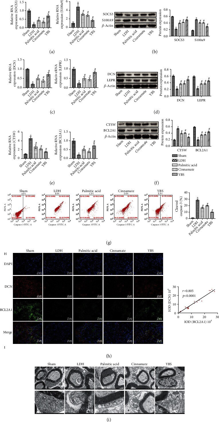
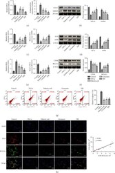
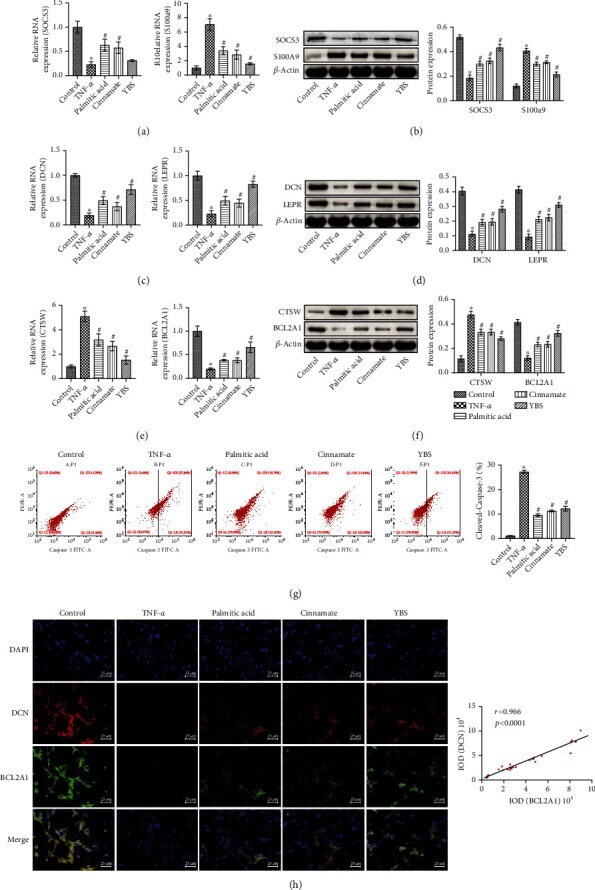
- Figure 4 YBS and its key compounds inhibited TNF- alpha -induced inflammation and apoptosis of DRG neuronal cells, and enhanced autophagy. (a) qRT-PCR was used to assess the expression of SOCS3 and S100A9 in cells. (b) Western blotting was used to detect the expression of SOCS3 and S100A9 in cells. (c) Detection of DCN and LEPR expression in cells by qRT-PCR. (d) The expression of DCN and LEPR in cells was detected by western blotting. (e) CTSW and BCL2A1 mRNA expression. (f) CTSW and BCL2A1 protein expression. (g) Flow cytometry to evaluate the expression of cleaved caspase-3 in cells. (h) The colocalization of DCN and BCL2A1 in cells was assessed by immunofluorescence. The magnification was 400x. Scale bar, 25 mu m. One- or two-way ANOVA was used for multiple group statistical analysis. * P < 0.05 vs. control group, # P < 0.05 vs TNF- alpha group.
- Submitted by
- Invitrogen Antibodies (provider)
- Main image
- Experimental details
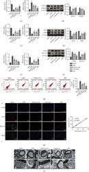
- The effect of YBS and its key compounds on the inflammation, apoptosis, and autophagy of DRG tissue in LDH rats. (a) qRT-PCR was applied to assess SOCS3 and S100A9 expression in tissues. (b) The expression of SOCS3 and S100A9 in the tissues was assessed by western blotting. (c) qRT-PCR was applied to detect the expression of DCN and LEPR in tissues. (d) Western blotting was used to measure the expression of DCN and LEPR in the tissue. (e) The expression of CTSW and BCL2A1 in tissues was used by qRT-PCR. (f) Western blotting was used to evaluate the expression of CTSW and BCL2A1 in tissues. (g) Flow cytometry was applied to detect the expression of cleaved caspase-3 in DRG neuron cells. (h) The colocalization of DCN and BCL2A1 in cells was detected by immunofluorescence. (i) TEM was used to observe the changes in cell microstructure in DRG tissue. The magnification was 400x. Scale bar, 25 mu m. One- or two-way ANOVA was used for multiple group statistical analysis. * P < 0.05 vs. sham group, # P < 0.05 vs. LDH group.