Explore
 Validate
 Learn
LearnA01064-3
antibody from Boster Biological Technology
Targeting: TNFRSF11A
CD265, FEO, LOH18CR1, PDB2, RANK
 Western blot
 ELISA
ELISAAntibody data
- Antibody Data
- Antigen structure
- References [1]
- Comments [0]
- Validations
- Western blot [1]
Submit
Validation data
Reference
Comment
Report error
- Product number
- A01064-3 - Provider product page

- Provider
- Boster Biological Technology
- Product name
- Anti-RANK/Tnfrsf11a Picoband™ Antibody
- Antibody type
- Polyclonal
- Description
- Rabbit IgG polyclonal antibody for RANK/Tnfrsf11a detection. Tested with WB, FCM, Direct ELISA in Mouse;Rat.
- Reactivity
- Mouse, Rat
- Host
- Rabbit
- Vial size
- 100μg/vial
- Concentration
- Add 0.2ml of distilled water will yield a concentration of 500ug/ml.
- Storage
- At -20°C for one year. After reconstitution, at 4°C for one month. It can also be aliquoted and stored frozen at -20°C for a longer time. Avoid repeated freezing and thawing.
- Handling
- Add 0.2ml of distilled water will yield a concentration of 500ug/ml.
Submitted references Effect of intervention initiation timing of pulsed electromagnetic field on ovariectomy-induced osteoporosis in rats.
Zhou J, Liao Y, Zeng Y, Xie H, Fu C, Li N
Bioelectromagnetics 2017 Sep;38(6):456-465
Bioelectromagnetics 2017 Sep;38(6):456-465
No comments: Submit comment
Supportive validation
- Submitted by
- Boster Biological Technology (provider)
- Main image
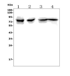
- Experimental details
- Western blot analysis of Tnfrsf11a using anti-Tnfrsf11a antibody (A01064-3). Electrophoresis was performed on a 5-20% SDS-PAGE gel at 70V (Stacking gel) / 90V (Resolving gel) for 2-3 hours. The sample well of each lane was loaded with 50ug of sample under reducing conditions. Lane 1: rat thymus tissue lysates, Lane 2: mouse thymus tissue lysates, Lane 3: mouse RAW264.7 whole cell lysates, Lane 4: mouse SP20 whole cell lysates. After Electrophoresis, proteins were transferred to a Nitrocellulose membrane at 150mA for 50-90 minutes. Blocked the membrane with 5% Non-fat Milk/ TBS for 1.5 hour at RT. The membrane was incubated with rabbit anti-Tnfrsf11a antigen affinity purified polyclonal antibody (Catalog # A01064-3) at 0.5 μg/mL overnight at 4°C, then washed with TBS-0.1%Tween 3 times with 5 minutes each and probed with a goat anti-rabbit IgG-HRP secondary antibody at a dilution of 1:5000 for 1.5 hour at RT. The signal is developed using an Enhanced Chemiluminescent detection (ECL) kit (Catalog # EK1002) with Tanon 5200 system. A specific band was detected for Tnfrsf11a at approximately 80KD. The expected band size for Tnfrsf11a is at 66KD.
- Additional image