Explore
 Validate
 Learn
Learn Western blot
 Immunohistochemistry
Antibody data
- Antibody Data
- Antigen structure
- References [3]
- Comments [0]
- Validations
- Western blot [3]
- Immunocytochemistry [1]
Submit
Validation data
Reference
Comment
Report error
- Product number
- GTX111637 - Provider product page

- Provider
- GeneTex
- Proper citation
- GeneTex Cat#GTX111637, RRID:AB_11167520
- Product name
- mAChR M3 antibody
- Antibody type
- Polyclonal
- Reactivity
- Human, Mouse
- Host
- Rabbit
Submitted references Single onabotulinumtoxinA 200U dose improved clinical symptoms but not urothelial dysfunction in neurogenic detrusor overactivity due to spinal cord injury.
Cell-Type-Specific Translation Profiling Reveals a Novel Strategy for Treating Fragile X Syndrome.
Suitability of Nicotinic Acetylcholine Receptor α7 and Muscarinic Acetylcholine Receptor 3 Antibodies for Immune Detection: Evaluation in Murine Skin.
Chen SF, Jiang YH, Kuo HC
Journal of the Formosan Medical Association = Taiwan yi zhi 2019 Jan;118(1 Pt 1):125-133
Journal of the Formosan Medical Association = Taiwan yi zhi 2019 Jan;118(1 Pt 1):125-133
Cell-Type-Specific Translation Profiling Reveals a Novel Strategy for Treating Fragile X Syndrome.
Thomson SR, Seo SS, Barnes SA, Louros SR, Muscas M, Dando O, Kirby C, Wyllie DJA, Hardingham GE, Kind PC, Osterweil EK
Neuron 2017 Aug 2;95(3):550-563.e5
Neuron 2017 Aug 2;95(3):550-563.e5
Suitability of Nicotinic Acetylcholine Receptor α7 and Muscarinic Acetylcholine Receptor 3 Antibodies for Immune Detection: Evaluation in Murine Skin.
Rommel FR, Raghavan B, Paddenberg R, Kummer W, Tumala S, Lochnit G, Gieler U, Peters EM
The journal of histochemistry and cytochemistry : official journal of the Histochemistry Society 2015 May;63(5):329-39
The journal of histochemistry and cytochemistry : official journal of the Histochemistry Society 2015 May;63(5):329-39
No comments: Submit comment
Supportive validation
- Submitted by
- GeneTex (provider)
- Main image
- Experimental details
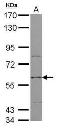
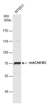
- Sample (30 ug of whole cell lysate) A: NT2D1 7.5% SDS PAGE GTX111637 diluted at 1:1000
- Validation comment
- WB

- Submitted by
- GeneTex (provider)
- Main image
- Experimental details
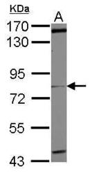
- Sample (50 ug of whole cell lysate) A: mouse brain 7.5% SDS PAGE GTX111637 diluted at 1:1000
- Validation comment
- WB

- Submitted by
- GeneTex (provider)
- Main image
- Experimental details
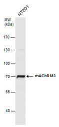
- mAChR M3 antibody detects mAChR M3 protein by western blot analysis. Whole cell extracts (30 ?g) was separated by 7.5% SDS-PAGE, and the membrane was blotted with mAChR M3 antibody (GTX111637) at a dilution of 1:1000. The HRP-conjugated anti-rabbit IgG antibody (GTX213110-01) was used to detect the primary antibody.
Supportive validation
- Submitted by
- GeneTex (provider)
- Main image
- Experimental details
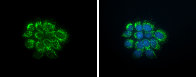
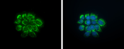
- mAChR M3 antibody detects mAChR M3 protein at cell membrane and cytoplasm by immunofluorescent analysis.Sample: A431 cells were fixed in ice-cold MeOH for 5 min.Green: mAChR M3 protein stained by mAChR M3 antibody (GTX111637) diluted at 1:500.Blue: Hoechst 33342 staining.