Explore
 Validate
 Learn
Learn Western blot
 Immunocytochemistry
Antibody data
- Antibody Data
- Antigen structure
- References [2]
- Comments [0]
- Validations
- Immunocytochemistry [4]
- Other assay [3]
Submit
Validation data
Reference
Comment
Report error
- Product number
- PA5-31773 - Provider product page

- Provider
- Invitrogen Antibodies
- Product name
- KIF23 Polyclonal Antibody
- Antibody type
- Polyclonal
- Antigen
- Recombinant full-length protein
- Description
- Recommended positive controls: HeLa. Predicted reactivity: Mouse (91%), Rat (90%), Bovine (93%). Store product as a concentrated solution. Centrifuge briefly prior to opening the vial.
- Reactivity
- Human
- Host
- Rabbit
- Isotype
- IgG
- Vial size
- 100 μL
- Concentration
- 0.8 mg/mL
- Storage
- Store at 4°C short term. For long term storage, store at -20°C, avoiding freeze/thaw cycles.
Submitted references Rab14/MACF2 complex regulates endosomal targeting during cytokinesis.
Knockdown of lncRNA PVT1 inhibits prostate cancer progression in vitro and in vivo by the suppression of KIF23 through stimulating miR-15a-5p.
Gibieža P, Peterman E, Hoffman HK, Van Engeleburg S, Skeberdis VA, Prekeris R
Molecular biology of the cell 2021 Apr 1;32(7):554-566
Molecular biology of the cell 2021 Apr 1;32(7):554-566
Knockdown of lncRNA PVT1 inhibits prostate cancer progression in vitro and in vivo by the suppression of KIF23 through stimulating miR-15a-5p.
Wu H, Tian X, Zhu C
Cancer cell international 2020;20:283
Cancer cell international 2020;20:283
No comments: Submit comment
Supportive validation
- Submitted by
- Invitrogen Antibodies (provider)
- Main image

- Experimental details
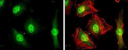
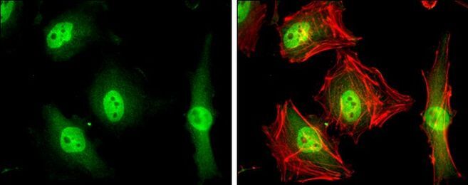
- Immunocytochemistry-Immunofluorescence analysis of KIF23 was performed in HeLa cells fixed in 4% paraformaldehyde at RT for 15 min. Green: KIF23 Polyclonal Antibody (Product # PA5-31773) diluted at 1:500. Red: phalloidin, a cytoskeleton marker.

- Submitted by
- Invitrogen Antibodies (provider)
- Main image

- Experimental details
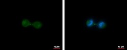
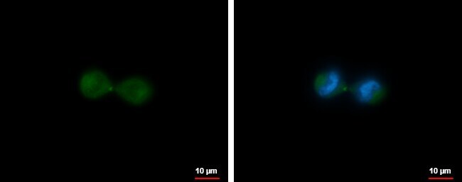
- KIF23 Polyclonal Antibody detects MKLP1 protein at midbody by immunofluorescent analysis. Sample: HeLa cells were fixed in 4% paraformaldehyde at RT for 15 min. Green: MKLP1 protein stained by KIF23 Polyclonal Antibody (Product # PA5-31773) diluted at 1:500. Blue: Hoechst 33342 staining. Scale bar = 10 µm.

- Submitted by
- Invitrogen Antibodies (provider)
- Main image
- Experimental details
- KIF23 Polyclonal Antibody detects MKLP1 protein at midbody by immunofluorescent analysis. Sample: HeLa cells were fixed in 4% paraformaldehyde at RT for 15 min. Green: MKLP1 protein stained by KIF23 Polyclonal Antibody (Product # PA5-31773) diluted at 1:500. Blue: Hoechst 33342 staining. Scale bar = 10 µm.
- Submitted by
- Invitrogen Antibodies (provider)
- Main image
- Experimental details
- Immunocytochemistry-Immunofluorescence analysis of KIF23 was performed in HeLa cells fixed in 4% paraformaldehyde at RT for 15 min. Green: KIF23 Polyclonal Antibody (Product # PA5-31773) diluted at 1:500. Red: phalloidin, a cytoskeleton marker.
Supportive validation
- Submitted by
- Invitrogen Antibodies (provider)
- Main image
- Experimental details
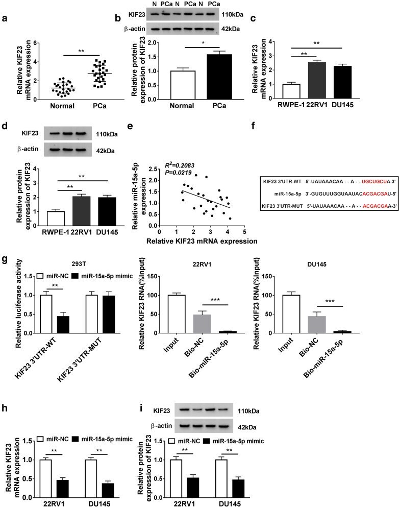
- Fig. 4 KIF23 was up-regulated in PCa tissues and cell lines and targeted by miR-15a-5p. a , b The expression of KIF23 at mRNA and protein levels in PCa tissues and normal tissues was measured by qRT-PCR and western blot, respectively. c , d The expression of KIF23 at mRNA and protein levels in PCa cells and normal cells was measured by qRT-PCR and western blot, respectively. e The expression of miR-15a-5p was negatively correlated with KIF23 expression. f The binding sites between miR-15a-5p and KIF23 3' UTR were forecasted by starBase v2.0. g The interaction between miR-15a-5p and KIF23 was confirmed by dual-luciferase reporter assay. h , i The expression of KIF23 was modulated by miR-15a-5p expression at both mRNA and protein levels. * P < 0.05, ** P < 0.01, *** P < 0.001
- Submitted by
- Invitrogen Antibodies (provider)
- Main image
- Experimental details
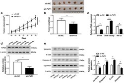
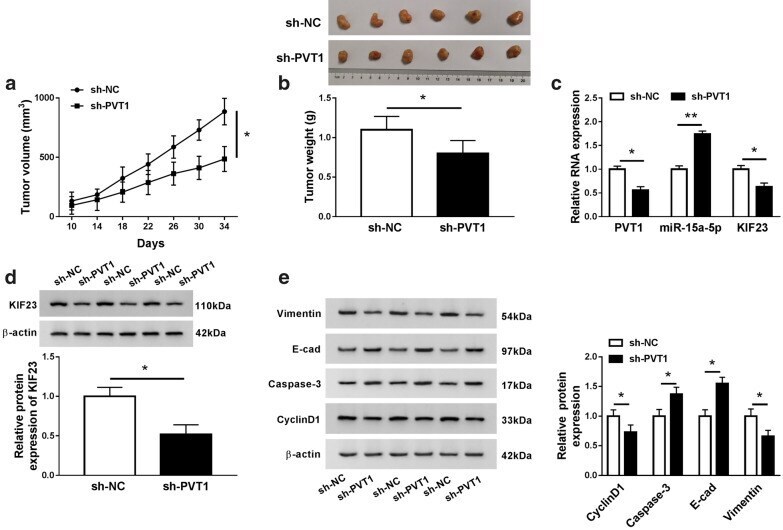
- Fig. 7 Knockdown of PVT1 impeded the PCa progression in vivo. a The tumor volume was calculated every 4 day after 10 day-inoculation. b The tumor weight was measured after 34 days. c The expression of PVT1, miR-15a-5p and KIF23, d the expression of KIF23 at the protein levels, e and the levels of Vimentin, E-cad, Caspase-3 and CyclinD1 were examined in removed tumor tissues using qRT-PCR or western blot. * P < 0.05, ** P < 0.01
- Submitted by
- Invitrogen Antibodies (provider)
- Main image
- Experimental details
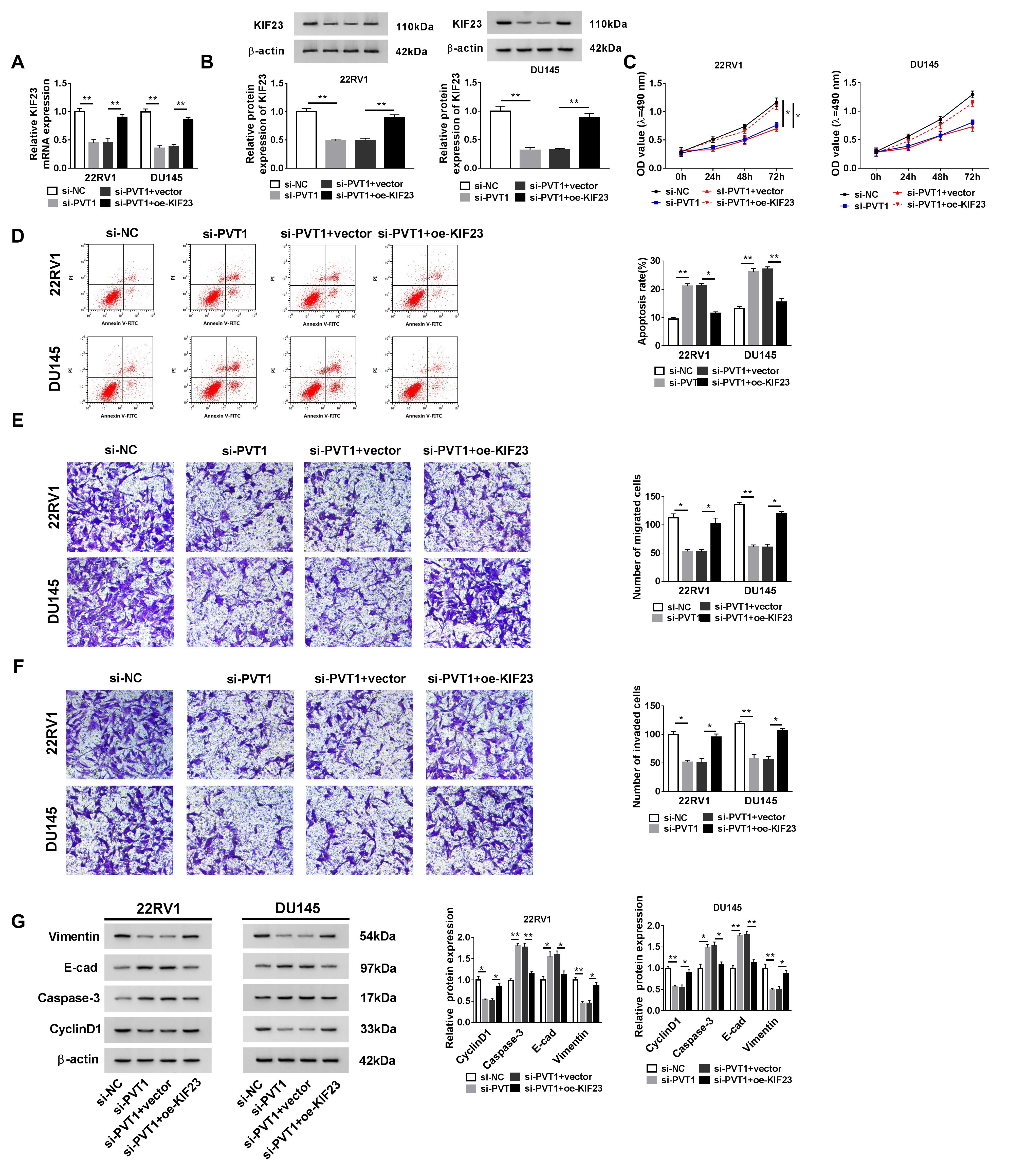
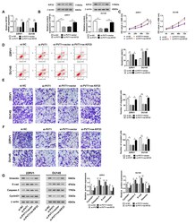
- Additional file 3: Figure S2. KIF23 overexpression rescued the effects of PVT1 knockdown. 22RV1 and DU145 cells were transfected with si-PVT1 or si-VT1 + oe-KIF23, with si-NC or or si-PVT1 + vector as the control. (A andB) The expression of KIF23 in these transfected cells was detected by qRT-PCR an western blot. (C) Cell proliferation was assessed by MTT assay. (D) Cell apoptosis was monitored by flow cytometry assay. (E and F) Cell migration and cell invasion in these transfected cells were investigated by transwell assay. (G) The expression of Vimentin, E-cad, Caspase-3 and CyclinD1 was quantified by western blot in these transfected cells. * P < 0.05, ** P < 0.01.