Explore
 Validate
 Learn
Learn Western blot
Antibody data
- Antibody Data
- Antigen structure
- References [4]
- Comments [0]
- Validations
- Western blot [4]
- Immunocytochemistry [1]
- Immunoprecipitation [1]
- Immunohistochemistry [1]
Submit
Validation data
Reference
Comment
Report error
- Product number
- GTX113850 - Provider product page

- Provider
- GeneTex
- Proper citation
- GeneTex Cat#GTX113850, RRID:AB_2037180
- Product name
- HNF1 alpha antibody [N1N3]
- Antibody type
- Polyclonal
- Reactivity
- Human, Mouse, Rat
- Host
- Rabbit
Submitted references Elevation of sensitivity to anticancer agents of human lung adenocarcinoma A549 cells by knockdown of claudin-2 expression in monolayer and spheroid culture models.
SATB1 is Down-regulated in Clear Cell Renal Cell Carcinoma and Correlates with miR-21-5p Overexpression and Poor Prognosis.
Effects of oolong tea on gene expression of gluconeogenic enzymes in the mouse liver and in rat hepatoma H4IIE cells.
Effects of (-)-epigallocatechin-3-O-gallate on expression of gluconeogenesis-related genes in the mouse duodenum.
Maruhashi R, Akizuki R, Sato T, Matsunaga T, Endo S, Yamaguchi M, Yamazaki Y, Sakai H, Ikari A
Biochimica et biophysica acta. Molecular cell research 2018 Mar;1865(3):470-479
Biochimica et biophysica acta. Molecular cell research 2018 Mar;1865(3):470-479
SATB1 is Down-regulated in Clear Cell Renal Cell Carcinoma and Correlates with miR-21-5p Overexpression and Poor Prognosis.
Kowalczyk AE, Krazinski BE, Godlewski J, Grzegrzolka J, Kiewisz J, Kwiatkowski P, Sliwinska-Jewsiewicka A, Dziegiel P, Kmiec Z
Cancer genomics & proteomics 2016 May-Jun;13(3):209-17
Cancer genomics & proteomics 2016 May-Jun;13(3):209-17
Effects of oolong tea on gene expression of gluconeogenic enzymes in the mouse liver and in rat hepatoma H4IIE cells.
Yasui K, Miyoshi N, Tababe H, Ishigami Y, Fukutomi R, Imai S, Isemura M
Journal of medicinal food 2011 Sep;14(9):930-8
Journal of medicinal food 2011 Sep;14(9):930-8
Effects of (-)-epigallocatechin-3-O-gallate on expression of gluconeogenesis-related genes in the mouse duodenum.
Yasui K, Tanabe H, Miyoshi N, Suzuki T, Goto S, Taguchi K, Ishigami Y, Paeng N, Fukutomi R, Imai S, Isemura M
Biomedical research (Tokyo, Japan) 2011 Oct;32(5):313-20
Biomedical research (Tokyo, Japan) 2011 Oct;32(5):313-20
No comments: Submit comment
Supportive validation
- Submitted by
- GeneTex (provider)
- Main image
- Experimental details
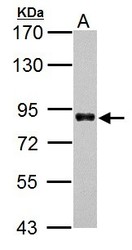
- HNF1 alpha antibody [N1N3] detects HNF1 alpha protein by western blot analysis.A. 30 ?g HepG2 whole cell lysate/extract 7.5% SDS-PAGEHNF1 alpha antibody [N1N3] (GTX113850) dilution: 1:5000 The HRP-conjugated anti-rabbit IgG antibody (GTX213110-01) was used to detect the primary antibody.
- Submitted by
- GeneTex (provider)
- Main image
- Experimental details
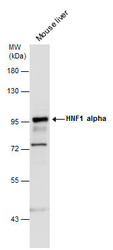
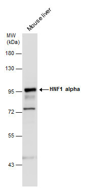
- HNF1 alpha antibody [N1N3] detects HNF1 alpha protein by western blot analysis. Mouse tissue extracts (50 ?g) was separated by 7.5% SDS-PAGE, and the membrane was blotted with HNF1 alpha antibody [N1N3] (GTX113850) diluted at 1:2000. The HRP-conjugated anti-rabbit IgG antibody (GTX213110-01) was used to detect the primary antibody.
- Submitted by
- GeneTex (provider)
- Main image
- Experimental details
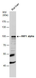
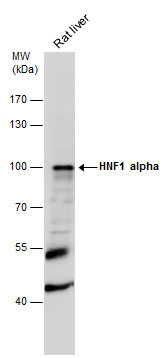
- HNF1 alpha antibody [N1N3] detects HNF1 alpha protein by western blot analysis. Rat tissue extracts (50 ?g) was separated by 7.5% SDS-PAGE, and the membrane was blotted with HNF1 alpha antibody [N1N3] (GTX113850) diluted at 1:1000. The HRP-conjugated anti-rabbit IgG antibody (GTX213110-01) was used to detect the primary antibody.
- Submitted by
- GeneTex (provider)
- Main image
- Experimental details
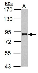
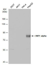
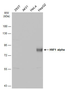
- Various whole cell extracts (30 ?g) were separated by 7.5% SDS-PAGE, and the membrane was blotted with HNF1 alpha antibody [N1N3] (GTX113850) diluted at 1:5000. The HRP-conjugated anti-rabbit IgG antibody (GTX213110-01) was used to detect the primary antibody.
Supportive validation
- Submitted by
- GeneTex (provider)
- Main image

- Experimental details
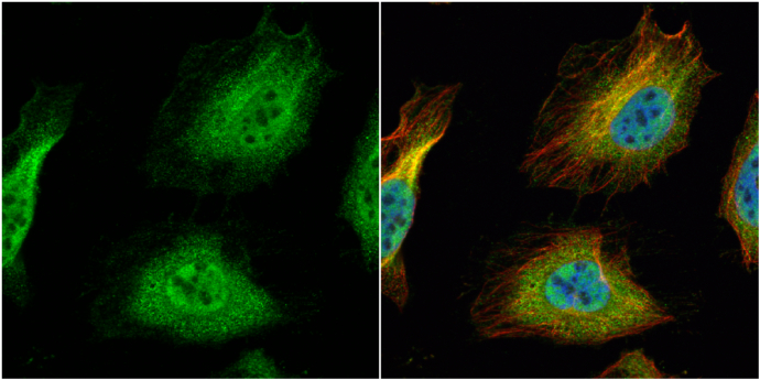
- HNF1 alpha antibody [N1N3] detects HNF1 alpha protein at cytoplasm and nucleus by immunofluorescent analysis.Sample: HeLa cells were fixed in 4% paraformaldehyde at RT for 15 min.Green: HNF1 alpha protein stained by HNF1 alpha antibody [N1N3] (GTX113850) diluted at 1:500.Red: alpha Tubulin, a cytoskeleton marker, stained by alpha Tubulin antibody [GT114] (GTX628802) diluted at 1:1000.Blue: Hoechst 33342 staining.
Supportive validation
- Submitted by
- GeneTex (provider)
- Main image
- Experimental details
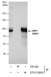
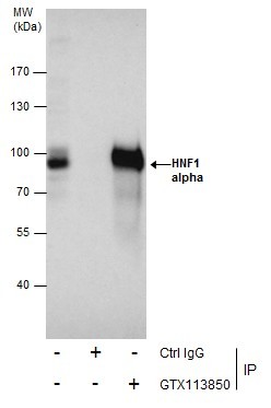
- Immunoprecipitation of HNF1 alpha protein from HepG2 whole cell extracts using 5 £gg of HNF1 alpha antibody [N1N3] (GTX113850).Western blot analysis was performed using HNF1 alpha antibody [N1N3] (GTX113850).EasyBlot anti-Rabbit IgG (GTX221666-01) was used as a secondary reagent.
Supportive validation
- Submitted by
- GeneTex (provider)
- Main image
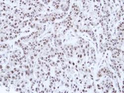
- Experimental details
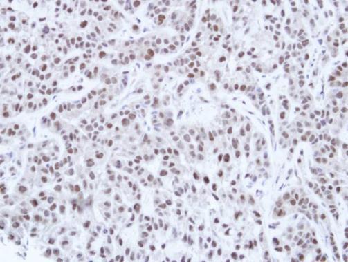
- Immunohistochemical analysis of paraffin-embedded A549 Xenograft, using HNF-1 alpha(GTX113850) antibody at 1:100 dilution.