Explore
 Validate
 Learn
Learn Western blot
 ELISA
ELISA Immunohistochemistry
Antibody data
- Antibody Data
- Antigen structure
- References [0]
- Comments [0]
- Validations
- Western blot [2]
- Immunohistochemistry [3]
Submit
Validation data
Reference
Comment
Report error
- Product number
- LS-C745375 - Provider product page

- Provider
- LSBio
- Product name
- SLC2A2 / GLUT2 Antibody (C-Terminus) LS-C745375
- Antibody type
- Polyclonal
- Description
- Affinity purified
- Reactivity
- Human, Mouse, Rat
- Host
- Rabbit
- Isotype
- IgG
- Storage
- Store vial at -20°C prior to opening. This product is stable for several weeks at 4°C as an undiluted liquid. Dilute only prior to immediate use. For extended storage aliquot contents and freeze at -20°C or below. Avoid freeze-thaw cycles.
No comments: Submit comment
Enhanced validation
- Submitted by
- LSBio (provider)
- Enhanced method
- Genetic validation
- Main image

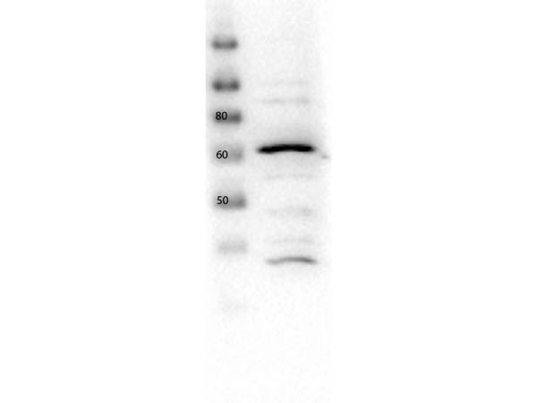
- Experimental details
- Western Blot of rabbit anti-Glut2 antibody. Lane 1: HEK293 lysate. Load: 10µg per lane. Primary antibody: Glut2 antibody at 1:1000 for overnight at 4°C. Secondary antibody: Peroxidase rabbit secondary antibody at 1:40,000 for 30 min at RT. Block: MB-070 overnight at 4°C. Predicted/Observed size: ~60 kDa.
- Submitted by
- LSBio (provider)
- Enhanced method
- Genetic validation
- Main image

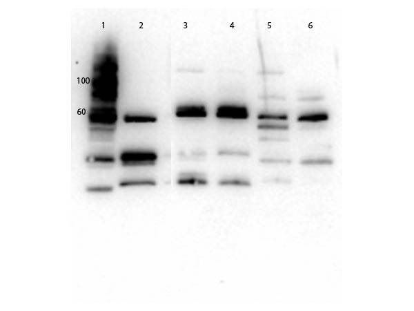
- Experimental details
- Western Blot of rabbit Anti-Glut2 Antibody. Lane 1: Molecular Weight. Lane 2: Mouse Kidney WCL. Lane 3: MEF WCL. Lane 4: 3T3 WCL. Lane 5: HeLa WCL. Lane 6: HEK293 WCL. Load: 20µg per pane. Primary antibody: 1µg/mL blocked with MB-073 overnight at 2-8°C. Secondary Antibody: Goat anti-Rabbit HRP 1:40,000 diluted with MB-073 for 30 minutes at RT. Expect: ~57.1 kDa.
Supportive validation
- Submitted by
- LSBio (provider)
- Enhanced method
- Genetic validation
- Main image

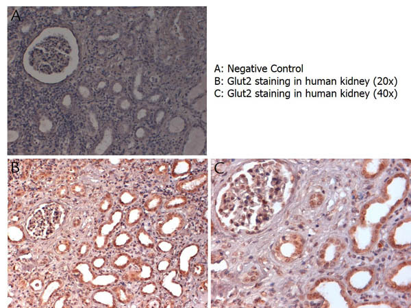
- Experimental details
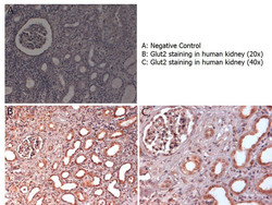
- Immunohistochemistry with anti-Glut2 antibody showing Glut2 staining in nucleus and cytoplasm of ductal epithelium and of renal glomeruli in human kidney at 20x and 40x (B & C). Formalin fixed/paraffin embedded sections were subjected to heat induced epitope retrieval (HIER) at pH 6.2 and then incubated with rabbit anti-mouse Glut2 antibody at 4.0 µg/ml for 60 minutes. The reaction was developed using MACH 1 universal HRP polymer detection system and visualized with 3’3-diamino-benzidine substrate (DAB).
- Submitted by
- LSBio (provider)
- Main image
- Experimental details
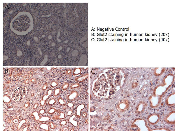
- Immunohistochemistry with anti-Glut2 antibody showing Glut2 staining in nucleus and cytoplasm of ductal epithelium and of renal glomeruli in human kidney at 20x and 40x (B & C). Formalin fixed/paraffin embedded sections were subjected to heat induced epitope retrieval (HIER) at pH 6.2 and then incubated with rabbit anti-mouse Glut2 antibody at 4.0 µg/ml for 60 minutes. The reaction was developed using MACH 1 universal HRP polymer detection system and visualized with 3’3-diamino-benzidine substrate (DAB).
- Submitted by
- LSBio (provider)
- Main image

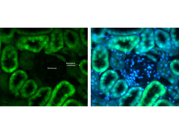
- Experimental details
- Immunohistochemistry of Anti-Glut2 Antibody. Tissue: mouse kidney. Antigen retrieval: Heat Induced, slides incubated in sodium citrate buffer for 1hr at 90°C. Primary: Rabbit Anti-Glut2 Antibody at 5µg/mL overnight. Blocking: 2% goat serum in TBST. Secondary: Alexa 488 at 1µg/mL for 2hrs at room temperature.