 Explore
 Validate
 Learn
Learn Western blot
 Immunohistochemistry
Antibody data
- Antibody Data
- Antigen structure
- References [2]
- Comments [0]
- Validations
- Immunohistochemistry [1]
Submit
Validation data
Reference
Comment
Report error
- Product number
- AF640 - Provider product page

- Provider
- Novus Biologicals
- Product name
- Goat Polyclonal EphA3 Antibody
- Antibody type
- Polyclonal
- Description
- Antigen Affinity-purified. Detects mouse EphA3 in direct ELISAs and Western blots. In direct ELISAs, approximately 5% cross-reactivity with recombinant mouse (rm) EphA4, rmEphA8, recombinant rat EphA5, and rmEphA6 is observed.
- Reactivity
- Mouse
- Host
- Goat
- Conjugate
- Unconjugated
- Isotype
- IgG
- Vial size
- 100 ug
- Concentration
- LYOPH
- Storage
- Use a manual defrost freezer and avoid repeated freeze-thaw cycles. 12 months from date of receipt, -20 to -70 degreesC as supplied. 1 month, 2 to 8 degreesC under sterile conditions after reconstitution. 6 months, -20 to -70 degreesC under sterile conditions after reconstitution.
Submitted references EphA3 null mutants do not demonstrate motor axon guidance defects.
EphA3 null mutants do not demonstrate motor axon guidance defects.
Vaidya A, Pniak A, Lemke G, Brown A
Molecular and cellular biology 2003 Nov;23(22):8092-8
Molecular and cellular biology 2003 Nov;23(22):8092-8
EphA3 null mutants do not demonstrate motor axon guidance defects.
Vaidya A, Pniak A, Lemke G, Brown A
Molecular and cellular biology 2003 Nov;23(22):8092-8
Molecular and cellular biology 2003 Nov;23(22):8092-8
No comments: Submit comment
Supportive validation
- Submitted by
- Novus Biologicals (provider)
- Main image
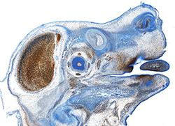
- Experimental details
- EphA3 in Mouse Embryo. EphA3 was detected in immersion fixed frozen sections of mouse embryo (13 d.p.c.) using Goat Anti-Mouse EphA3 Antigen Affinity-purified Polyclonal Antibody (Catalog # AF640) at 1.7 µg/mL overnight at 4 °C. Tissue was stained using the Anti-Goat HRP-DAB Cell & Tissue Staining Kit (brown; Catalog # CTS008) and counterstained with hematoxylin (blue). Specific staining was localized to developing brain. View our protocol for Chromogenic IHC Staining of Frozen Tissue Sections.
