Explore
 Validate
 Learn
Learn Western blot
Antibody data
- Antibody Data
- Antigen structure
- References [0]
- Comments [0]
- Validations
- Western blot [3]
- Immunohistochemistry [1]
- Flow cytometry [1]
Submit
Validation data
Reference
Comment
Report error
- Product number
- PA5-111771 - Provider product page

- Provider
- Invitrogen Antibodies
- Product name
- EphA3 (extracellular) Polyclonal Antibody
- Antibody type
- Polyclonal
- Antigen
- Synthetic peptide
- Description
- Applications Tested: This 61D3 antibody has been pre-titrated and tested by flow cytometric analysis of normal peripheral blood cells. This can be used at 5 µL (0.5 µg) per test. A test is defined as the amount (µg) of antibody that will stain a cell sample in a final volume of 100 µL. Cell number should be determined empirically but can range from 10^5 to 10^8 cells/test.
- Reactivity
- Human, Mouse, Rat
- Host
- Rabbit
- Isotype
- IgG
- Vial size
- 50 µL
- Concentration
- 0.8 mg/mL
- Storage
- -20°C
No comments: Submit comment
Supportive validation
- Submitted by
- Invitrogen Antibodies (provider)
- Main image
- Experimental details
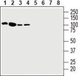
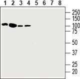
- Western Blot analysis of EphA3 was performed in human Jurkat T-cell leukemia cell line lysate (lanes 1 and 5), human K562 chronic myelogenous leukemia cell line lysate (lanes 2 and 6), human Malme-3M melanoma cell line lysate (lanes 3 and 7) and human HT-29 colorectal adenocarcinoma cell line lysate (lanes 4 and 8). Lane 1-4: EphA3 (extracellular) Antibody (Product # PA5-111771) at a dilution of 1:200. Lane 5-8: EphA3 (extracellular) Antibody preincubated with the negative control antigen.
- Submitted by
- Invitrogen Antibodies (provider)
- Main image
- Experimental details
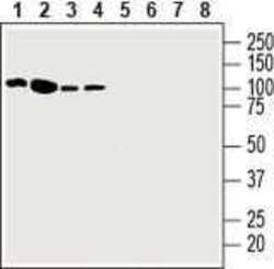
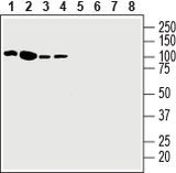
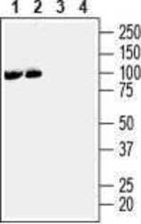
- Western Blot analysis of EphA3 was performed in mouse (lanes 1 and 3) and rat (lanes 2 and 4) brain membranes. Lane 1,2: EphA3 (extracellular) Antibody (Product # PA5-111771) at a dilution of 1:200. Lane 3,4: EphA3 (extracellular) Antibody preincubated with the negative control antigen.

Supportive validation
- Submitted by
- Invitrogen Antibodies (provider)
- Main image
- Experimental details
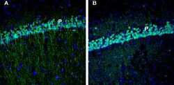
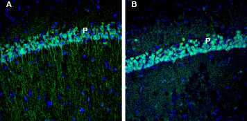
- Immunohistochemistry analysis of EphA3 in perfusion-fixed, frozen rat and mouse hippocampus tissue sections using EphA3 (extracellular) Antibody (Product # PA5-111771) at a dilution of 1:200, followed by goat anti-rabbit-AlexaFluor-488. A) EphA3 staining (green) in rat hippocampal CA1 region is detected in the pyramidal layer (P). B) In mouse hippocampal CA1 region, EphA3 immunoreactivity (green) is observed in the pyramidal layer (P). Cell nuclei are stained with DAPI (blue).
Supportive validation
- Submitted by
- Invitrogen Antibodies (provider)
- Main image

- Experimental details
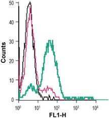
- Flow Cytometry analysis of EphA3 in live intact human Jurkat T-cell leukemia cells. Black: Cells. Pink: Cells and goat-anti-rabbit-FITC. Green: Cells, EphA3 (extracellular) Antibody (Product # PA5-111771), and goat-anti-rabbit-FITC.