Explore
 Validate
 Learn
Learn Western blot
 ELISA
ELISAAntibody data
- Antibody Data
- Antigen structure
- References [1]
- Comments [0]
- Validations
- Western blot [1]
Submit
Validation data
Reference
Comment
Report error
- Product number
- A00578 - Provider product page

- Provider
- Boster Biological Technology
- Product name
- Anti-Eph receptor A2/EPHA2 Antibody Picoband™
- Antibody type
- Polyclonal
- Description
- Polyclonal antibody for ECK/EPHA2 detection. Host: Rabbit.Size: 100μg/vial. Tested applications: WB, FCM, ICC/IF Direct ELISA. Reactive species: Human. ECK/EPHA2 information: Subcellular Localization: Cell membrane; Tissue Specificity: Expressed in brain and glioma tissue and glioma cell lines (at protein level). Expressed most highly in tissues that contain a high proportion of epithelial cells, e.g. skin, intestine, lung, and ovary.
- Reactivity
- Human, Mouse, Rat
- Host
- Rabbit
- Vial size
- 100μg/vial
- Concentration
- 0.5-1mg/ml, actual concentration vary by lot. Use suggested dilution ratio to decide dilution procedure.
- Storage
- At -20°C for one year. After reconstitution, at 4°C for one month. It can also be aliquoted and stored frozen at -20°C for a longer time. Avoid repeated freezing and thawing.
- Handling
- Add 0.2ml of distilled water will yield a concentration of 500ug/ml.
Submitted references Effect of ephrin-A1/EphA2 on invasion of trophoblastic cells.
Yang Y, Min J
Journal of Huazhong University of Science and Technology. Medical sciences = Hua zhong ke ji da xue xue bao. Yi xue Ying De wen ban = Huazhong keji daxue xuebao. Yixue Yingdewen ban 2011 Dec;31(6):824-827
Journal of Huazhong University of Science and Technology. Medical sciences = Hua zhong ke ji da xue xue bao. Yi xue Ying De wen ban = Huazhong keji daxue xuebao. Yixue Yingdewen ban 2011 Dec;31(6):824-827
No comments: Submit comment
Supportive validation
- Submitted by
- Boster Biological Technology (provider)
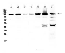
- Main image
- Experimental details
- Western blot analysis of Eph receptor A2 using anti-Eph receptor A2 antibody (A00578). Electrophoresis was performed on a 5-20% SDS-PAGE gel at 70V (Stacking gel) / 90V (Resolving gel) for 2-3 hours. The sample well of each lane was loaded with 50ug of sample under reducing conditions. Lane 1: human Hela cell lysate,Lane 2: human U-87MG cell lysate,Lane 3: human SHG-44 cell lysate,Lane 4: human COLO-320 cell lysate,Lane 5: human SK-OV-3 cell lysate,Lane 6: human A549 cell lysate,Lane 7: mouse HEPA1-6 cell lysate. After Electrophoresis, proteins were transferred to a Nitrocellulose membrane at 150mA for 50-90 minutes. Blocked the membrane with 5% Non-fat Milk/ TBS for 1.5 hour at RT. The membrane was incubated with rabbit anti-Eph receptor A2 antigen affinity purified polyclonal antibody (Catalog # A00578) at 0.5 μg/mL overnight at 4°C, then washed with TBS-0.1%Tween 3 times with 5 minutes each and probed with a goat anti-rabbit IgG-HRP secondary antibody at a dilution of 1:10000 for 1.5 hour at RT. The signal is developed using an Enhanced Chemiluminescent detection (ECL) kit (Catalog # EK1002) with Tanon 5200 system. A specific band was detected for Eph receptor A2 at approximately 125KD. The expected band size for Eph receptor A2 is at 108KD.
- Additional image