Explore
 Validate
 Learn
Learn Western blot
Antibody data
- Antibody Data
- Antigen structure
- References [6]
- Comments [0]
- Validations
- Western blot [1]
- Immunohistochemistry [1]
- Flow cytometry [1]
Submit
Validation data
Reference
Comment
Report error
- Product number
- AF3035 - Provider product page

- Provider
- R&D Systems
- Product name
- Human EphA2 Antibody
- Antibody type
- Polyclonal
- Description
- Antigen Affinity-purified. Detects human EphA2 in direct ELISAs and Western blots. In direct ELISAs, less than 45% cross-reactivity with recombinant mouse EphA2 is observed and less than 1% cross-reactivity with recombinant human (rh) EphA1, rhEphA3, rhEphA4, rhEphA5, rhEphA6, rhEphA7 and rhEphA10 is observed.
- Reactivity
- Human
- Host
- Goat
- Conjugate
- Unconjugated
- Antigen sequence
P29317- Isotype
- IgG
- Vial size
- 100 ug
- Concentration
- LYOPH
- Storage
- Use a manual defrost freezer and avoid repeated freeze-thaw cycles. 12 months from date of receipt, -20 to -70 °C as supplied. 1 month, 2 to 8 °C under sterile conditions after reconstitution. 6 months, -20 to -70 °C under sterile conditions after reconstitution.
Submitted references Stemness underpinning all steps of human colorectal cancer defines the core of effective therapeutic strategies.
Engineered nanointerfaces for microfluidic isolation and molecular profiling of tumor-specific extracellular vesicles.
Protein kinase A can block EphA2 receptor-mediated cell repulsion by increasing EphA2 S897 phosphorylation.
Alteration of the EphA2/Ephrin-A signaling axis in psoriatic epidermis.
EphA2 cleavage by MT1-MMP triggers single cancer cell invasion via homotypic cell repulsion.
A novel extracellular Hsp90 mediated co-receptor function for LRP1 regulates EphA2 dependent glioblastoma cell invasion.
Visioli A, Giani F, Trivieri N, Pracella R, Miccinilli E, Cariglia MG, Palumbo O, Arleo A, Dezi F, Copetti M, Cajola L, Restelli S, Papa V, Sciuto A, Latiano TP, Carella M, Amadori D, Gallerani G, Ricci R, Alfieri S, Pesole G, Vescovi AL, Binda E
EBioMedicine 2019 Jun;44:346-360
EBioMedicine 2019 Jun;44:346-360
Engineered nanointerfaces for microfluidic isolation and molecular profiling of tumor-specific extracellular vesicles.
Reátegui E, van der Vos KE, Lai CP, Zeinali M, Atai NA, Aldikacti B, Floyd FP Jr, H Khankhel A, Thapar V, Hochberg FH, Sequist LV, Nahed BV, S Carter B, Toner M, Balaj L, T Ting D, Breakefield XO, Stott SL
Nature communications 2018 Jan 12;9(1):175
Nature communications 2018 Jan 12;9(1):175
Protein kinase A can block EphA2 receptor-mediated cell repulsion by increasing EphA2 S897 phosphorylation.
Barquilla A, Lamberto I, Noberini R, Heynen-Genel S, Brill LM, Pasquale EB
Molecular biology of the cell 2016 Sep 1;27(17):2757-70
Molecular biology of the cell 2016 Sep 1;27(17):2757-70
Alteration of the EphA2/Ephrin-A signaling axis in psoriatic epidermis.
Gordon K, Kochkodan JJ, Blatt H, Lin SY, Kaplan N, Johnston A, Swindell WR, Hoover P, Schlosser BJ, Elder JT, Gudjonsson JE, Getsios S
The Journal of investigative dermatology 2013 Mar;133(3):712-722
The Journal of investigative dermatology 2013 Mar;133(3):712-722
EphA2 cleavage by MT1-MMP triggers single cancer cell invasion via homotypic cell repulsion.
Sugiyama N, Gucciardo E, Tatti O, Varjosalo M, Hyytiäinen M, Gstaiger M, Lehti K
The Journal of cell biology 2013 Apr 29;201(3):467-84
The Journal of cell biology 2013 Apr 29;201(3):467-84
A novel extracellular Hsp90 mediated co-receptor function for LRP1 regulates EphA2 dependent glioblastoma cell invasion.
Gopal U, Bohonowych JE, Lema-Tome C, Liu A, Garrett-Mayer E, Wang B, Isaacs JS
PloS one 2011 Mar 8;6(3):e17649
PloS one 2011 Mar 8;6(3):e17649
No comments: Submit comment
Supportive validation
- Submitted by
- R&D Systems (provider)
- Main image
- Experimental details
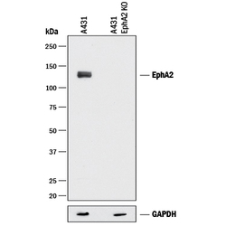
- Detection of Human EphA2 by Western Blot. Western blot shows lysates of A431 human epithelial carcinoma parental cell line and EphA2 knock out (KO) A431 cell line. PVDF membrane was probed with 1 µg/mL of Goat Anti-Human EphA2 Antigen Affinity-purified Polyclonal Antibody (Catalog # AF3035) followed by HRP-conjugated Anti-Rabbit IgG Secondary Antibody (Catalog # HAF008). A specific band was detected for EphA2 at approximately 110 kDa (as indicated), but not detectable in the knockout A431 cell line. GAPDH (Catalog # MAB5718) is shown as a loading control. This experiment was conducted under reducing conditions and using Immunoblot Buffer Group 1.
Supportive validation
- Submitted by
- R&D Systems (provider)
- Main image
- Experimental details
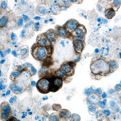
- EphA2 in Human Ovarian Cancer Tissue. EphA2 was detected in immersion fixed paraffin-embedded sections of human ovarian cancer tissue using Goat Anti-Human EphA2 Antigen Affinity-purified Polyclonal Antibody (Catalog # AF3035) at 5 µg/mL overnight at 4 °C. Tissue was stained using the Anti-Goat HRP-DAB Cell & Tissue Staining Kit (brown; Catalog # CTS008) and counterstained with hematoxylin (blue). Specific labeling was localized to the plasma membrane of cancer cells. View our protocol for Chromogenic IHC Staining of Paraffin-embedded Tissue Sections.

Supportive validation
- Submitted by
- R&D Systems (provider)
- Main image

- Experimental details
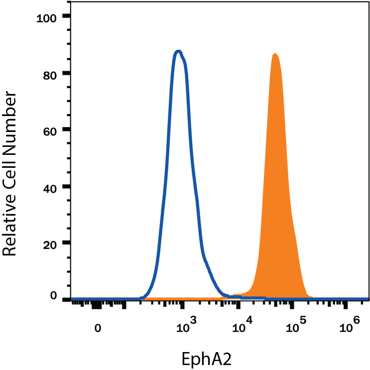
- Detection of EphA2 in A431 Human Cell Line by Flow Cytometry. A431 human epithelial carcinoma cell line was stained with Goat Anti-Human EphA2 Antigen Affinity-purified Polyclonal Antibody (Catalog # AF3035, filled histogram) or isotype control antibody (Catalog # AB-108-C, open histogram), followed by Phycoerythrin-conjugated Anti-Goat IgG Secondary Antibody (Catalog # F0107). View our protocol for Staining Membrane-associated Proteins.