 Explore
 Validate
 Learn
Learn Immunocytochemistry
Antibody data
- Antibody Data
- Antigen structure
- References [4]
- Comments [0]
- Validations
- Immunocytochemistry [2]
- Flow cytometry [1]
Submit
Validation data
Reference
Comment
Report error
- Product number
- MAB3035 - Provider product page

- Provider
- R&D Systems
- Product name
- Human EphA2 Antibody
- Antibody type
- Monoclonal
- Description
- Protein A or G purified from hybridoma culture supernatant. Detects human EphA2 in direct ELISAs and Western blots. In direct ELISAs, no cross-reactivity with recombinant mouse EphA4, A5, A6, A7, A8, or recombinant rat EphB1 is observed.
- Reactivity
- Human
- Host
- Mouse
- Conjugate
- Unconjugated
- Antigen sequence
P29317- Isotype
- IgG
- Antibody clone number
- 371805
- Vial size
- 100 ug
- Concentration
- LYOPH
- Storage
- Use a manual defrost freezer and avoid repeated freeze-thaw cycles. 12 months from date of receipt, -20 to -70 °C as supplied. 1 month, 2 to 8 °C under sterile conditions after reconstitution. 6 months, -20 to -70 °C under sterile conditions after reconstitution.
Submitted references Engineering of monobody conjugates for human EphA2-specific optical imaging.
Isolation and Characterization of a Monobody with a Fibronectin Domain III Scaffold That Specifically Binds EphA2.
Quantitative assessment of antibody internalization with novel monoclonal antibodies against Alexa fluorophores.
Plk5, a polo box domain-only protein with specific roles in neuron differentiation and glioblastoma suppression.
Kim MA, Yoon HS, Park SH, Kim DY, Pyo A, Kim HS, Min JJ, Hong Y
PloS one 2017;12(7):e0180786
PloS one 2017;12(7):e0180786
Isolation and Characterization of a Monobody with a Fibronectin Domain III Scaffold That Specifically Binds EphA2.
Park SH, Park S, Kim DY, Pyo A, Kimura RH, Sathirachinda A, Choy HE, Min JJ, Gambhir SS, Hong Y
PloS one 2015;10(7):e0132976
PloS one 2015;10(7):e0132976
Quantitative assessment of antibody internalization with novel monoclonal antibodies against Alexa fluorophores.
Liao-Chan S, Daine-Matsuoka B, Heald N, Wong T, Lin T, Cai AG, Lai M, D'Alessio JA, Theunissen JW
PloS one 2015;10(4):e0124708
PloS one 2015;10(4):e0124708
Plk5, a polo box domain-only protein with specific roles in neuron differentiation and glioblastoma suppression.
de Cárcer G, Escobar B, Higuero AM, García L, Ansón A, Pérez G, Mollejo M, Manning G, Meléndez B, Abad-Rodríguez J, Malumbres M
Molecular and cellular biology 2011 Mar;31(6):1225-39
Molecular and cellular biology 2011 Mar;31(6):1225-39
No comments: Submit comment
Supportive validation
- Submitted by
- R&D Systems (provider)
- Main image
- Experimental details
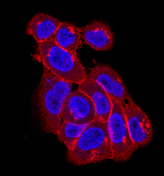
- EphA2 in A431 Human Cell Line. EphA2 was detected in immersion fixed A431 human epithelial carcinoma cell line using Mouse Anti-Human EphA2 Monoclonal Antibody (Catalog # MAB3035) at 10 µg/mL for 3 hours at room temperature. Cells were stained using the NorthernLights™ 557-conjugated Anti-Mouse IgG Secondary Antibody (red; Catalog # NL007) and counterstained with DAPI(blue). Specific staining was localized to the cell surface. View our protocol for Fluorescent ICC Staining of Cells on Coverslips.

- Submitted by
- R&D Systems (provider)
- Main image
- Experimental details
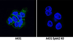
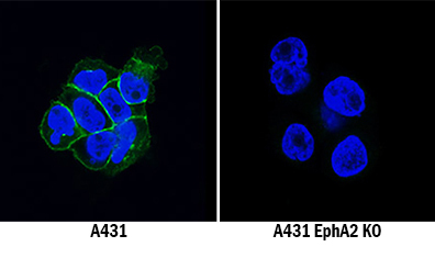
- EphA2 Specificity is Shown by Immunocytochemistry in Knockout Cell Line. EphA2 was detected in immersion fixed A431 human epithelial carcinoma cell line but is not detected in EphA2 knockout (KO) A431 Human Cell Line cell line using Mouse Anti-Human EphA2 Monoclonal Antibody (Catalog # MAB3035) at 5 µg/mL for 3 hours at room temperature. Cells were stained using the NorthernLights 493-conjugated Anti-Mouse IgG Secondary Antibody (green; Catalog # NL009) and counterstained with DAPI (blue). Specific staining was localized to cell membranes. View our protocol for Fluorescent ICC Staining of Cells on Coverslips.
Supportive validation
- Submitted by
- R&D Systems (provider)
- Main image
- Experimental details
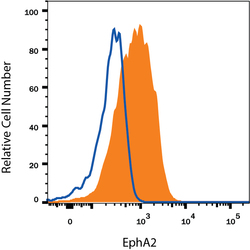
- Detection of EphA2 in A431 Human Cell Line by Flow Cytometry. A431 human epithelial carcinoma cell line was stained with Mouse Anti-Human EphA2 Monoclonal Antibody (Catalog # MAB3035, filled histogram) or isotype control antibody (Catalog # MAB003, open histogram), followed by Phycoerythrin-conjugated Anti-Mouse IgG Secondary Antibody (Catalog # F0102B). View our protocol for Staining Membrane-associated Proteins.
