Explore
 Validate
 Learn
Learn Western blot
 ELISA
ELISAAntibody data
- Antibody Data
- Antigen structure
- References [4]
- Comments [0]
- Validations
- Western blot [1]
- Immunocytochemistry [1]
- Flow cytometry [1]
Submit
Validation data
Reference
Comment
Report error
- Product number
- ABIN179724 - Provider product page

- Provider
- antibodies-online
- Proper citation
- Antibodies-Online Cat#ABIN179724, RRID:AB_10953878
- Product name
- anti-EPH Receptor A2 (EPHA2) antibody
- Antibody type
- Monoclonal
- Antigen
- genetic immunisation with cDNA encoding human EphA2
- Description
- Protein G
- Reactivity
- Human
- Isotype
- IgG
- Antibody clone number
- Kalpha-5H5
- Vial size
- 100 μg
- Concentration
- 2 mg/mL
- Storage
- short term: 2°C - 8°C, long term: -20°C
- Handling
- Avoid repeated freezing and thawing.
Submitted references EphA2 receptor tyrosine kinase as a promising target for cancer therapeutics.
Eph receptor tyrosine kinases in tumor and tumor microenvironment.
Diverse roles for the Eph family of receptor tyrosine kinases in carcinogenesis.
cDNA cloning and characterization of eck, an epithelial cell receptor protein-tyrosine kinase in the eph/elk family of protein kinases.
Ireton RC, Chen J
Current cancer drug targets 2005 May;5(3):149-57
Current cancer drug targets 2005 May;5(3):149-57
Eph receptor tyrosine kinases in tumor and tumor microenvironment.
Brantley-Sieders D, Schmidt S, Parker M, Chen J
Current pharmaceutical design 2004;10(27):3431-42
Current pharmaceutical design 2004;10(27):3431-42
Diverse roles for the Eph family of receptor tyrosine kinases in carcinogenesis.
Nakamoto M, Bergemann AD
Microscopy research and technique 2002 Oct 1;59(1):58-67
Microscopy research and technique 2002 Oct 1;59(1):58-67
cDNA cloning and characterization of eck, an epithelial cell receptor protein-tyrosine kinase in the eph/elk family of protein kinases.
Lindberg RA, Hunter T
Molecular and cellular biology 1990 Dec;10(12):6316-24
Molecular and cellular biology 1990 Dec;10(12):6316-24
No comments: Submit comment
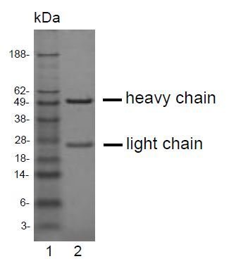
Supportive validation
- Submitted by
- antibodies-online (provider)
- Main image
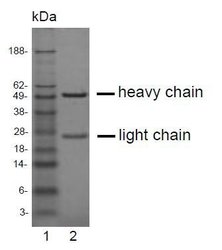
- Experimental details
- SDS-PAGE analysis of purified K?-5H5 monoclonal antibody. Lane 1: molecular weight marker, Lane 2: 2 ?g of purified K?-5H5 antibody. Proteins were separated by SDS-PAGE and stained with RAPID StainTM Reagent.
Supportive validation
- Submitted by
- antibodies-online (provider)
- Main image
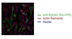
- Experimental details
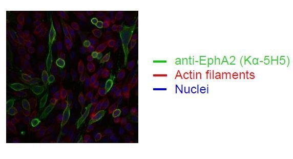
- Spectral Confocal Microscopy of CHO cells using K?-5H5. CHO cells were transiently transfected with an expression vector encoding EphA2. Binding of K?-5H5 was visualized with a FITC-conjugated secondary antibody (green). Actin filaments are labeled with Alexa Fluor-555 Phalloidin (red). Cell nuclei are stained with DAPI (blue).
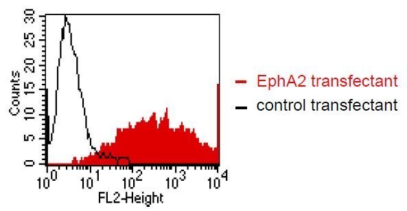
Supportive validation
- Submitted by
- antibodies-online (provider)
- Main image
- Experimental details
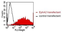
- FACS analysis of BOSC23 cells using K?-5H5. BOSC23 cells were transiently trans-fected with an expression vector encoding either EphA2 (red curve) or an irrelevant protein (control transfectant). Binding of K?-5H5 was detected with a PE-conjugated secondary antibody. A positive signal was obtained only with EphA2 transfected cells.