 Explore
 Validate
 Learn
Learn Western blot
Antibody data
- Antibody Data
- Antigen structure
- References [0]
- Comments [0]
- Validations
- Western blot [2]
- Immunocytochemistry [2]
Submit
Validation data
Reference
Comment
Report error
- Product number
- ABIN2508748 - Provider product page

- Provider
- antibodies-online
- Product name
- anti-Melanoma Antigen Family D, 1 (MAGED1) (Center) antibody
- Antibody type
- Monoclonal
- Antigen
- This MAGED1 antibody is generated from a mouse immunized with a KLH conjugated synthetic peptide between 20-224 amino acids from the Central region of human MAGED1.Antigen type: Recombinant Protein
- Description
- This antibody is purified through a protein G column, followed by dialysis against PBS.
- Reactivity
- Human
- Host
- Mouse
- Epitope
- Center
- Isotype
- IgG
- Vial size
- 200 μL
- Storage
- Maintain refrigerated at 2-8°C for up to 6 months. For long term storage store at -20°C in small aliquots to prevent freeze-thaw cycles.
No comments: Submit comment
Supportive validation
- Submitted by
- antibodies-online (provider)
- Main image

- Experimental details
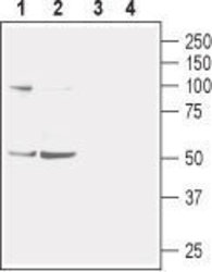
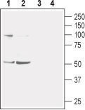
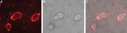
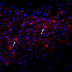
- Western blot analysis of mouse (lanes 1 and 3) and rat (lanes 2 and 4) brain membranes: 1-2. Anti-Nicotinic Acetylcholine Receptor ?2 (extracellular) antibody (ABIN2511196), (1:400). 3-4. Anti-Nicotinic Acetylcholine Receptor ?2 (extracellular) antibody, preincubated with the control peptide antigen. Western blot analysis of human SH-SY5Y neuroblastoma cell lysate: 1. Anti-Nicotinic Acetylcholine Receptor ?2 (extracellular) antibody (ABIN2511196), (1:200). 2. Anti-Nicotinic Acetylcholine Receptor ?2 (extracellular) antibody, preincubated with the control peptide antigen. Expression of nAChR?2 in rat deep cerebellar nucleus Immunohistochemical staining of immersion-fixed, free floating rat brain frozen sections using Anti-Nicotinic Acetylcholine Receptor ?2 (extracellular) antibody (ABIN2511196), (1:100). Staining reveals expression of nAChR?2 (red) in cells with neuronal outline (arrows point at some examples) in the red nucleus. DAPI is used as the counterstain (blue). Expression of Nicotinic Acetylcholine Receptor ?2 in rat PC12 pheochromocytoma cells Immunocytochemical staining of live intact rat PC12 pheochromocytoma cells. A. Extracellular staining of live cells with Anti-Nicotinic Acetylcholine Receptor ?2 (extracellular) antibody (ABIN2511196), (1:50), followed by goat anti-rabbit-AlexaFluor-594 (red). B. Live image of the cells. C. Merge of the two images.

- Submitted by
- antibodies-online (provider)
- Main image
- Experimental details
- Western blot analysis of mouse (lanes 1 and 3) and rat (lanes 2 and 4) brain membranes: 1-2. Anti-Nicotinic Acetylcholine Receptor ?2 (extracellular) antibody (ABIN2511196), (1:400). 3-4. Anti-Nicotinic Acetylcholine Receptor ?2 (extracellular) antibody, preincubated with the control peptide antigen. Western blot analysis of human SH-SY5Y neuroblastoma cell lysate: 1. Anti-Nicotinic Acetylcholine Receptor ?2 (extracellular) antibody (ABIN2511196), (1:200). 2. Anti-Nicotinic Acetylcholine Receptor ?2 (extracellular) antibody, preincubated with the control peptide antigen. Expression of nAChR?2 in rat deep cerebellar nucleus Immunohistochemical staining of immersion-fixed, free floating rat brain frozen sections using Anti-Nicotinic Acetylcholine Receptor ?2 (extracellular) antibody (ABIN2511196), (1:100). Staining reveals expression of nAChR?2 (red) in cells with neuronal outline (arrows point at some examples) in the red nucleus. DAPI is used as the counterstain (blue). Expression of Nicotinic Acetylcholine Receptor ?2 in rat PC12 pheochromocytoma cells Immunocytochemical staining of live intact rat PC12 pheochromocytoma cells. A. Extracellular staining of live cells with Anti-Nicotinic Acetylcholine Receptor ?2 (extracellular) antibody (ABIN2511196), (1:50), followed by goat anti-rabbit-AlexaFluor-594 (red). B. Live image of the cells. C. Merge of the two images.
Supportive validation
- Submitted by
- antibodies-online (provider)
- Main image

- Experimental details
- Image(s): Immunofluorescence
- Submitted by
- antibodies-online (provider)
- Main image
- Experimental details
- Image(s): Immunofluorescence
