Explore
 Validate
 Learn
Learn Western blot
 Immunohistochemistry
Antibody data
- Antibody Data
- Antigen structure
- References [0]
- Comments [0]
- Validations
- Western blot [2]
- Immunocytochemistry [1]
- Immunohistochemistry [6]
Submit
Validation data
Reference
Comment
Report error
- Product number
- HPA017929 - Provider product page

- Provider
- Atlas Antibodies
- Proper citation
- Atlas Antibodies Cat#HPA017929, RRID:AB_1858770
- Product name
- Anti-VRK1
- Antibody type
- Polyclonal
- Reactivity
- Human
- Host
- Rabbit
- Conjugate
- Unconjugated
- Antigen sequence
LPWEDNLKDPKYVRDSKIRYRENIASLMDKCFPEK
NKPGEIAKYMETVKLLDYTEKPLYENLRDILLQGL
KAIGSKDDGKLDLSVVENGGLKAKTITKKRKKEIE
ESKEPGVEDTEWSNTQTEEAIQTRSRTR- Isotype
- IgG
- Vial size
- 100 µl
- Storage
- Store at +4°C for short term storage. Long time storage is recommended at -20°C.
No comments: Submit comment
Supportive validation
- Submitted by
- Atlas Antibodies (provider)
- Enhanced method
- Genetic validation
- Main image
- Experimental details
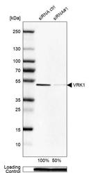
- Western blot analysis in Rh30 cells transfected with control siRNA, target specific siRNA probe #1, using Anti-VRK1 antibody. Remaining relative intensity is presented. Loading control: Anti-PPIB.

- Submitted by
- Atlas Antibodies (provider)
- Main image
- Experimental details
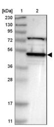
- Lane 1: Marker [kDa] 230, 130, 95, 72, 56, 36, 28, 17, 11Lane 2: Human cell line RT-4

Supportive validation
- Submitted by
- Atlas Antibodies (provider)
- Main image
- Experimental details
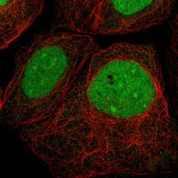
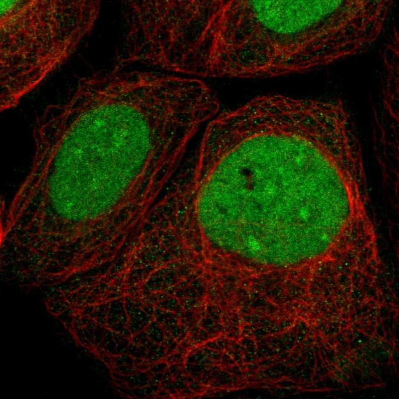
- Immunofluorescent staining of human cell line A-431 shows localization to nucleus & nucleoli.
- Sample type
- HUMAN
Enhanced validation
Supportive validation
- Submitted by
- Atlas Antibodies (provider)
- Enhanced method
- Orthogonal validation
- Main image
- Experimental details
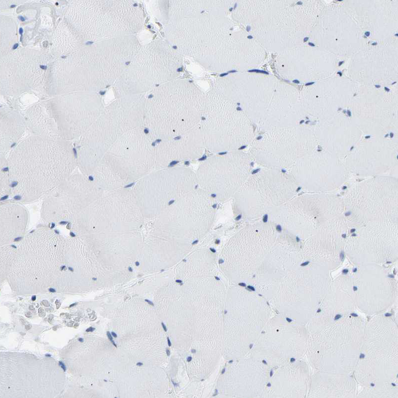
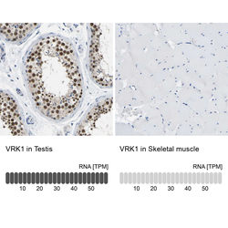
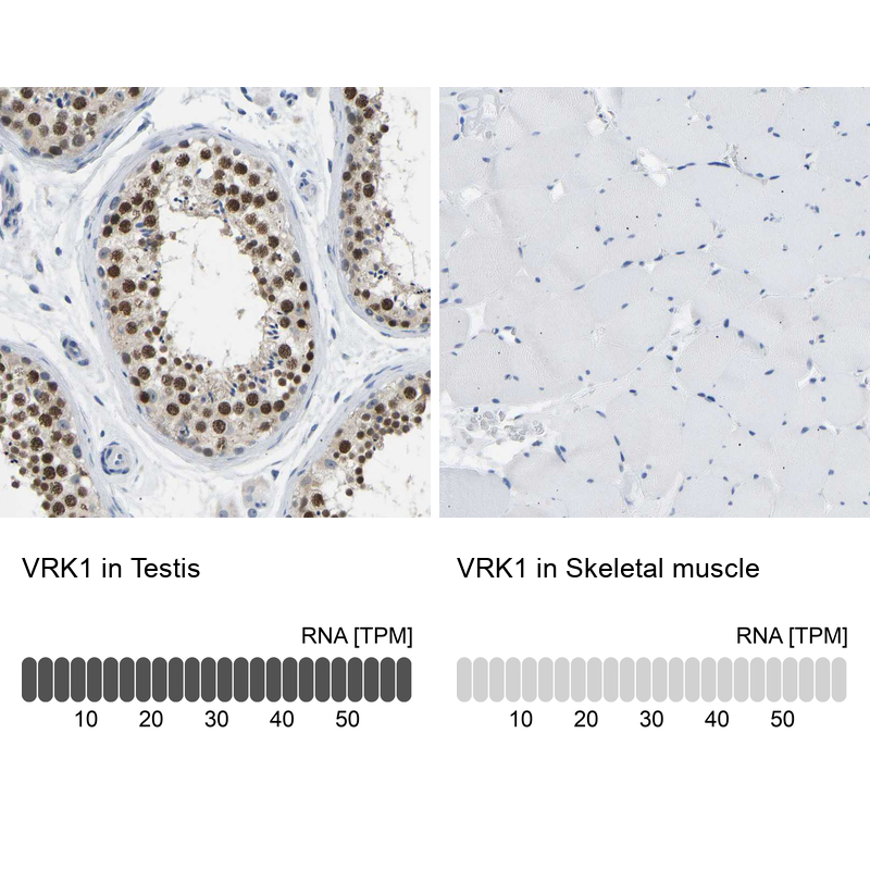
- Immunohistochemistry analysis in human testis and skeletal muscle tissues using HPA017929 antibody. Corresponding VRK1 RNA-seq data are presented for the same tissues.
- Sample type
- HUMAN
Supportive validation
- Submitted by
- Atlas Antibodies (provider)
- Main image

- Experimental details
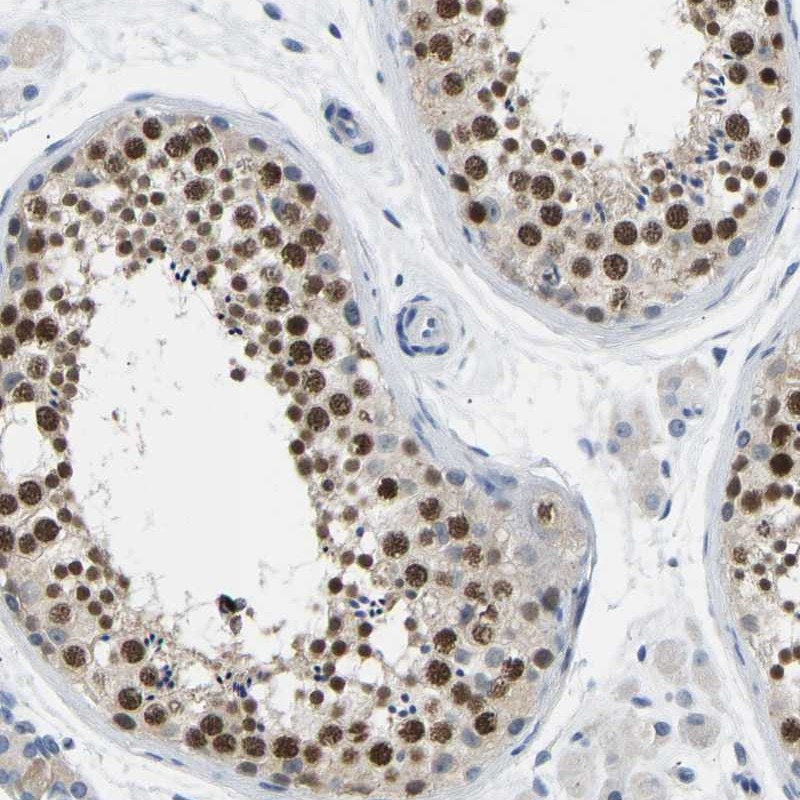
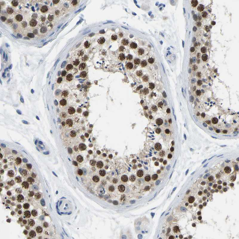
- Immunohistochemical staining of human testis shows strong nuclear positivity in cells of seminiferus ducts.
- Sample type
- HUMAN
- Submitted by
- Atlas Antibodies (provider)
- Main image
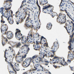
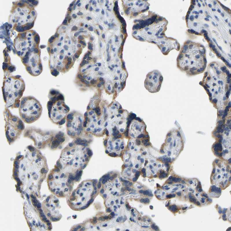
- Experimental details
- Immunohistochemical staining of human placenta shows moderate cytoplasmic positivity in trophoblastic cells.
- Sample type
- HUMAN
- Submitted by
- Atlas Antibodies (provider)
- Main image

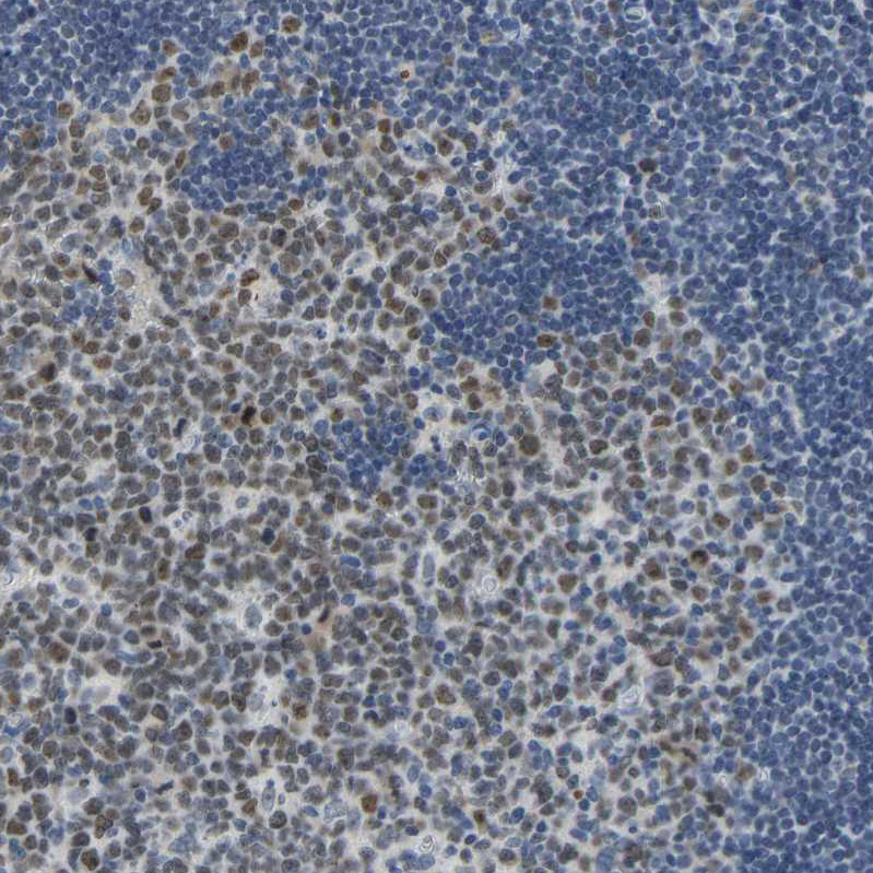
- Experimental details
- Immunohistochemical staining of human tonsil shows moderate nuclear positivity in germinal center cells.
- Sample type
- HUMAN
- Submitted by
- Atlas Antibodies (provider)
- Main image
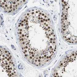
- Experimental details
- Immunohistochemical staining of human testis shows strong nuclear positivity in cells in seminiferous ducts.
- Sample type
- HUMAN
- Submitted by
- Atlas Antibodies (provider)
- Main image

- Experimental details
- Immunohistochemical staining of human skeletal muscle shows no positivity in myocytes as expected.
- Sample type
- HUMAN