Explore
 Validate
 Learn
Learn Western blot
Antibody data
- Antibody Data
- Antigen structure
- References [0]
- Comments [0]
- Validations
- Western blot [1]
- Immunoprecipitation [1]
- Immunohistochemistry [11]
Submit
Validation data
Reference
Comment
Report error
- Product number
- GTX84224 - Provider product page

- Provider
- GeneTex
- Proper citation
- GeneTex Cat#GTX84224, RRID:AB_10729745
- Product name
- LDHA antibody [2B11]
- Antibody type
- Monoclonal
- Reactivity
- Human
- Host
- Mouse
- Storage
- For short-term storage, store at 4¢XC or aliquot into working amounts and store at -20¢XC. For long-term storage, store at -70¢XC (aliquotted). Avoid repeated freeze-thaw cycles.
No comments: Submit comment
Supportive validation
- Submitted by
- GeneTex (provider)
- Main image
- Experimental details
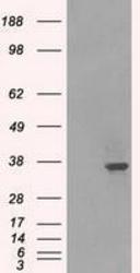
- HEK293T cells were transfected with the pCMV6-ENTRY control (Left lane) or pCMV6-ENTRY LDHA (Right lane) cDNA for 48 hrs and lysed. Equivalent amounts of cell lysates (5 ug per lane) were separated by SDS-PAGE and immunoblotted with anti-LDHA.

Supportive validation
- Submitted by
- GeneTex (provider)
- Main image
- Experimental details
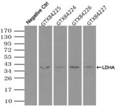
- Immunoprecipitation (IP) of LDHA by using TrueMab monoclonal anti-LDHA antibodies (Negative control: IP without adding anti-LDHA antibody.). For each experiment, 500ul of DDK tagged LDHA overexpression lysates (at 1:5 dilution with HEK293T lysate), 2ug of anti-LDHA antibody and 20ul (0.1mg) of goat anti-mouse conjugated magnetic beads were mixed and incubated overnight. After extensive wash to remove any non-specific binding, the immuno-precipitated products were analyzed with rabbit anti-DDK polyclonal antibody.

Supportive validation
- Submitted by
- GeneTex (provider)
- Main image
- Experimental details
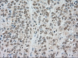
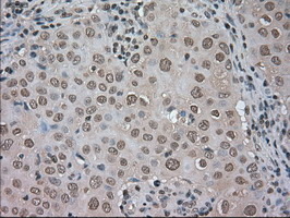
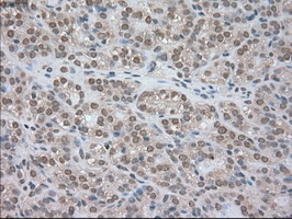
- Immunohistochemical staining of paraffin-embedded adenocarcinoma of human breast tissue using anti-LDHA mouse monoclonal antibody. (GTX84224)
- Submitted by
- GeneTex (provider)
- Main image

- Experimental details
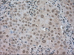
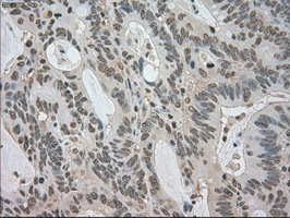
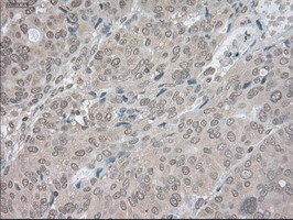
- Immunohistochemical staining of paraffin-embedded adenocarcinoma of human colon tissue using anti-LDHA mouse monoclonal antibody. (GTX84224)
- Submitted by
- GeneTex (provider)
- Main image
- Experimental details
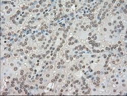
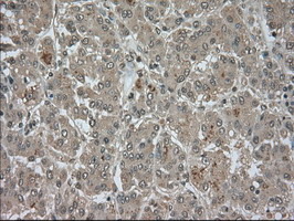
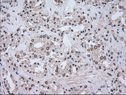
- Immunohistochemical staining of paraffin-embedded carcinoma of human kidney tissue using anti-LDHA mouse monoclonal antibody. (GTX84224)
- Submitted by
- GeneTex (provider)
- Main image

- Experimental details
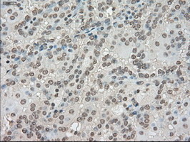
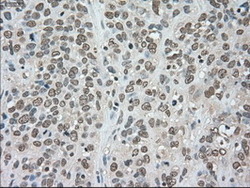
- Immunohistochemical staining of paraffin-embedded carcinoma of human liver tissue using anti-LDHA mouse monoclonal antibody. (GTX84224)
- Submitted by
- GeneTex (provider)
- Main image

- Experimental details
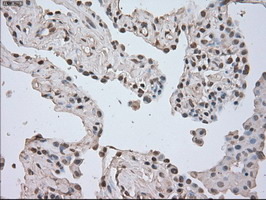
- Immunohistochemical staining of paraffin-embedded carcinoma of human lung tissue using anti-LDHA mouse monoclonal antibody. (GTX84224)
- Submitted by
- GeneTex (provider)
- Main image
- Experimental details
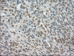
- Immunohistochemical staining of paraffin-embedded adenocarcinoma of human ovary tissue using anti-LDHA mouse monoclonal antibody. (GTX84224)

- Submitted by
- GeneTex (provider)
- Main image

- Experimental details
- Immunohistochemical staining of paraffin-embedded carcinoma of human pancreas tissue using anti-LDHA mouse monoclonal antibody. (GTX84224)
- Submitted by
- GeneTex (provider)
- Main image

- Experimental details
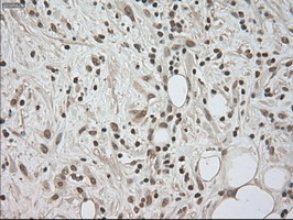
- Immunohistochemical staining of paraffin-embedded carcinoma of human thyroid tissue using anti-LDHA mouse monoclonal antibody. (GTX84224)
- Submitted by
- GeneTex (provider)
- Main image

- Experimental details
- Immunohistochemical staining of paraffin-embedded adenocarcinoma of human endometrium tissue using anti-LDHA mouse monoclonal antibody. (GTX84224)
- Submitted by
- GeneTex (provider)
- Main image
- Experimental details
- Immunohistochemical staining of paraffin-embedded carcinoma of human prostate tissue using anti-LDHA mouse monoclonal antibody. (GTX84224)

- Submitted by
- GeneTex (provider)
- Main image
- Experimental details
- Immunohistochemical staining of paraffin-embedded carcinoma of human bladder tissue using anti-LDHA mouse monoclonal antibody. (GTX84224)