 Explore
 Validate
 Learn
Learn Western blot
 Immunoprecipitation
Antibody data
- Antibody Data
- Antigen structure
- References [0]
- Comments [0]
- Validations
- Western blot [3]
- Immunocytochemistry [1]
- Immunohistochemistry [1]
Submit
Validation data
Reference
Comment
Report error
- Product number
- PA5-19754 - Provider product page

- Provider
- Invitrogen Antibodies
- Product name
- LDHA Polyclonal Antibody
- Antibody type
- Polyclonal
- Antigen
- Synthetic peptide
- Description
- Heat mediated antigen retrieval recommended prior to tissue staining. This antibody is predicted to react with mouse, rat, rabbit, chimpanzee and chinese hamster based on sequence homology.
- Reactivity
- Human
- Host
- Rabbit
- Isotype
- IgG
- Vial size
- 100 µg
- Concentration
- 0.9 mg/mL
- Storage
- Store at 4°C short term. For long term storage, store at -20°C, avoiding freeze/thaw cycles.
No comments: Submit comment
Supportive validation
- Submitted by
- Invitrogen Antibodies (provider)
- Main image

- Experimental details
- Western blot analysis of HeLa Whole Cell Lysate using Product # PA5-19754, Proteasome 19S S7 primary antibody at a dilution of 1 µg/mL (lane 1). Staining of Jurkat Whole Cell Lysate at a dilution of 1 µg/mL (lane 2). Blot treated with a secondary HRP-conjugated Goat polyclonal anti-Rabbit antibody was used at a dilution of 1:3000.

- Submitted by
- Invitrogen Antibodies (provider)
- Main image

- Experimental details
- Knockdown of LDHA was achieved by transfecting PC-3 with LDHA specific siRNAs (Silencer® select Product # S350, S351). Western blot analysis (Fig. a) was performed using Whole Cell Extract-WCL from the LDHA knockdown cells (lane 3), non-targeting scrambled siRNA transfected cells (lane 2) and untransfected cells (lane 1). The blot was probed with LDHA Polyclonal Antibody (Product # PA5-19754, 1 ug/ml ) and Goat anti-Rabbit IgG (H+L) Superclonal™ Recombinant Secondary Antibody, HRP (Product # A27036, 1:4000 dilution). Densitometric analysis of this western blot is shown in histogram (Fig. b). Decrease in signal upon siRNA mediated knock down confirms that antibody is specific to LDHA.

- Submitted by
- Invitrogen Antibodies (provider)
- Main image

- Experimental details
- Western blot was performed using Anti-LDHA Polyclonal Antibody(Product # PA5-19754) and a 35kDa band corresponding to LDHA was observed across cell lines and tissue extracts tested. A non-specific band around 50kDa has also been picked up in tissues extracts. Whole Cell Extract-WCL (30 µg lysate) of PC-3 (Lane 1), DU 145 (Lane 2), PANC-1 (Lane 3), MCF7 (Lane 4), MDA-MB-231 (Lane 5) and tissue extracts of Mouse Skeletal Muscle (Lane 6) and Rat Skeletal Muscle (Lane 7) were electrophoresed using NuPAGE™ 4-12% Bis-Tris Protein Gel (Product # NP0322BOX). Resolved proteins were then transferred onto a Nitrocellulose membrane (Product # IB23001) by iBlot® 2 Dry Blotting System (Product # IB21001). The blot was probed with the primary antibody (1 ug/ml) and detected by chemiluminescence with Goat anti-Rabbit IgG (H+L) Superclonal™ Recombinant Secondary Antibody, HRP (Product # A27036, 1:4000 dilution) using the iBright FL 1000 (Product # A32752). Chemiluminescent detection was performed using Novex® ECL Chemiluminescent Substrate Reagent Kit (Product # WP20005).

Supportive validation
- Submitted by
- Invitrogen Antibodies (provider)
- Main image
- Experimental details
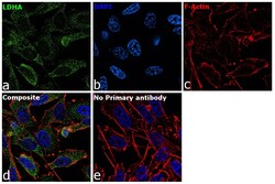
- Immunofluorescence analysis of LDHA was performed using 70% confluent log phase PC-3 cells. The cells were fixed with 4% paraformaldehyde for 10 minutes, permeabilized with 0.1% Triton™ X-100 for 15 minutes, and blocked with 2% BSA for 45 minutes at room temperature. The cells were labeled with LDHA Polyclonal Antibody (Product # PA5-19754) at 2.5 µg/mL in 0.1% BSA, incubated at 4 degree celsius overnight and then labeled with Goat anti-Rabbit IgG (H+L) Highly Cross-Adsorbed Secondary Antibody, Alexa Fluor Plus 488 (Product # A32731), (1:2000), for 45 minutes at room temperature (Panel a: Green). Nuclei (Panel b:Blue) were stained with ProLong™ Diamond Antifade Mountant with DAPI (Product # P36962). F-actin (Panel c: Red) was stained with Rhodamine Phalloidin (Product # R415, 1:300). Panel d represents the merged image showing cytoplasmic localization. Panel e represents control cells with no primary antibody to assess background. The images were captured at 60X magnification.

Supportive validation
- Submitted by
- Invitrogen Antibodies (provider)
- Main image
- Experimental details
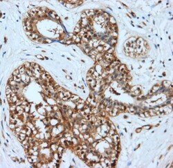
- Immunohistochemical (formalin-fixed, paraffin-embedded) staining of Human Breast Carcinoma tissue using Product # PA5-19754, anti-LDHA antibody. Primary antibody was used at a concentration of 1 µg/mL and exposed for 15 mins at room temp. The sample was pretreated using heat mediated antigen retrieval with Sodium Citrate Buffer (pH6/20mins). The detection method was a HRP conjugated polymer, DAB chromogen and the sample was counterstained with haematoxylin and mounted with DPX.
