Explore
 Validate
 Learn
Learn Western blot
 Immunohistochemistry
Antibody data
- Antibody Data
- Antigen structure
- References [2]
- Comments [0]
- Validations
- Immunohistochemistry [1]
- Flow cytometry [2]
- Other assay [3]
Submit
Validation data
Reference
Comment
Report error
- Product number
- PA5-26531 - Provider product page

- Provider
- Invitrogen Antibodies
- Product name
- LDHA Polyclonal Antibody
- Antibody type
- Polyclonal
- Antigen
- Synthetic peptide
- Description
- This antibody is predicted to react with bovine, porcine, non-human primate and rat based on sequence homology.
- Reactivity
- Human
- Host
- Rabbit
- Isotype
- IgG
- Vial size
- 400 μL
- Concentration
- 0.34 mg/mL
- Storage
- Store at 4°C short term. For long term storage, store at -20°C, avoiding freeze/thaw cycles.
Submitted references CircRNA circYY1 (hsa_circ_0101187) Modulates Cell Glycolysis and Malignancy Through Regulating YY1 Expression by Sponging miR-769-3p in Breast Cancer.
Lactate dehydrogenase expression modulates longevity and neurodegeneration in Drosophila melanogaster.
Zhang X, Li J, Feng Q
Cancer management and research 2021;13:1145-1158
Cancer management and research 2021;13:1145-1158
Lactate dehydrogenase expression modulates longevity and neurodegeneration in Drosophila melanogaster.
Long DM, Frame AK, Reardon PN, Cumming RC, Hendrix DA, Kretzschmar D, Giebultowicz JM
Aging 2020 Jun 2;12(11):10041-10058
Aging 2020 Jun 2;12(11):10041-10058
No comments: Submit comment
Supportive validation
- Submitted by
- Invitrogen Antibodies (provider)
- Main image

- Experimental details
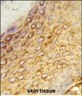
- Immunohistochemistry analysis of LDHA in formalin-fixed and paraffin-embedded human skin. Samples were incubated with LDHA polyclonal antibody (Product # PA5-26531) which was peroxidase-conjugated to the secondary antibody, followed by DAB staining. This data demonstrates the use of this antibody for immunohistochemistry; clinical relevance has not been evaluated.
Supportive validation
- Submitted by
- Invitrogen Antibodies (provider)
- Main image
- Experimental details
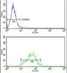
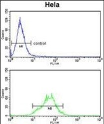
- Flow cytometry analysis of HeLa cells using a LDHA polyclonal antibody (Product # PA5-26531) (bottom) compared to a negative control cell (top) at a dilution of 1:10-50, followed by a FITC-conjugated goat anti-rabbit antibody
- Submitted by
- Invitrogen Antibodies (provider)
- Main image
- Experimental details
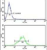
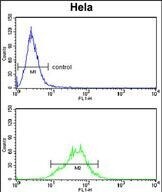
- Flow cytometry of LDHA in Hela cells (bottom histogram). Samples were incubated with LDHA polyclonal antibody (Product # PA5-26531) followed by FITC-conjugated goat-anti-rabbit secondary antibody. Negative control cell (top histogram).
Supportive validation
- Submitted by
- Invitrogen Antibodies (provider)
- Main image
- Experimental details
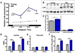
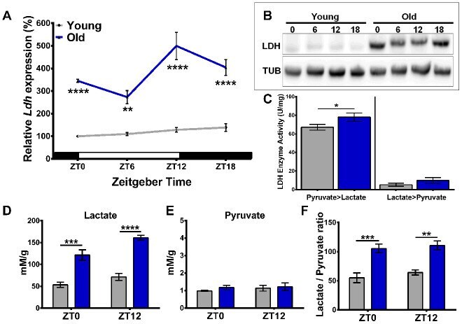
- Figure 1 Age-related increase in Ldh expression is associated with elevated LDH protein levels, enzyme activity, and lactate concentration. ( A ) Profile of Ldh mRNA expression in the heads of young (5-days-old) and old (55-days-old) w 1118 flies measured by qRT-PCR. Compared to young, old flies have increased Ldh mRNA levels at each time point with a peak at ZT12. Values are averages of 4 biorepeats reported as a percentage of expression relative to young at ZT0 set to 100%. ( B ) Representative western blot of LDH protein levels in young and old fly heads with tubulin as a loading control. ( C ) Graph showing LDH enzyme activity for both the pyruvate to lactate and lactate to pyruvate reactions in the heads of young (grey bars) and old (blue bars) flies. Enzymatic activity of LDH is higher in the heads of old flies compared to young for the pyruvate to lactate reaction (*p
- Submitted by
- Invitrogen Antibodies (provider)
- Main image
- Experimental details
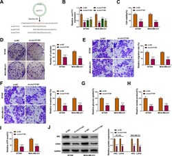
- Figure 2 Influence of circYY1 inhibition on the malignancy and glycolysis of BC cells. ( A ) Schematic diagram showed 3 siRNAs targeting the back-spliced sequence of circYY1. ( B ) QRT-PCR verified the knockdown efficiency of circYY1 in BT549 and MDA-MB-231 cells. ( C - F ) Influence of circYY1 inhibition on viability, colony formation, migration, and invasion of BT549 and MDA-MB-231 cells was assessed by MTT assay ( C ), colony formation assay ( D ), or transwell assay ( E and F ). ( G - I ) Impacts of circYY1 knockdown on glucose uptake, lactate production, and ATP release in BT549 and MDA-MB-231 cells were determined with corresponding kits. ( J ) Effects of circYY1 silencing on the protein levels of HK2 and LDHA in BT549 and MDA-MB-231 cells were analyzed using Western blotting. ** P < 0.01 and *** P < 0.001.

- Submitted by
- Invitrogen Antibodies (provider)
- Main image

- Experimental details
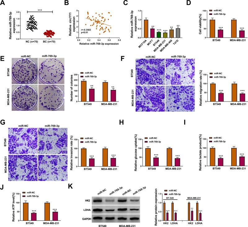
- Figure 4 Effects of miR-769-3p overexpression on the malignancy and glycolysis of BC cells. ( A ) Expression pattern of miR-769-3p in BC tissues and neighboring normal tissues was validated by qRT-PCR. ( B ) Analysis of the correlation between miR-769-3p and circYY1 in BC tissues by Pearson's correlation analysis. ( C ) Analysis of the expression of miR-769-3p in BC cells and MCF10A cells by qRT-PCR. ( D - K ) BT549 and MDA-MB-231 cells were transfected with miR-769-3p mimic and miR-NC. ( D - G ) The viability, colony formation, migration, and invasion of BT549 and MDA-MB-231 cells were analyzed by MTT assay ( D ), colony formation assay ( E ), or transwell assay ( F and G ). ( H - J ) Corresponding kits were used to analyze the levels of glucose uptake, lactate production, and ATP release in BT549 and MDA-MB-231 cells. ( K ) Western blotting presented the protein levels of HK2 and LDHA in BT549 and MDA-MB-231 cells. ** P < 0.01 and *** P < 0.001.