Explore
 Validate
 Learn
Learn Western blot
 Immunohistochemistry
Antibody data
- Antibody Data
- Antigen structure
- References [6]
- Comments [0]
- Validations
- Immunohistochemistry [1]
Submit
Validation data
Reference
Comment
Report error
- Product number
- HPA017888 - Provider product page

- Provider
- Atlas Antibodies
- Proper citation
- Atlas Antibodies Cat#HPA017888, RRID:AB_1858355
- Product name
- Anti-TNNT2
- Antibody type
- Polyclonal
- Description
- Polyclonal Antibody against Human TNNT2, Gene description: troponin T type 2 (cardiac), Alternative Gene Names: CMD1D, CMH2, CMPD2, Validated applications: IHC, WB, Uniprot ID: P45379, Storage: Store at +4°C for short term storage. Long time storage is recommended at -20°C.
- Reactivity
- Human
- Host
- Rabbit
- Conjugate
- Unconjugated
- Isotype
- IgG
- Vial size
- 100 µl
- Concentration
- 0.2 mg/ml
- Storage
- Store at +4°C for short term storage. Long time storage is recommended at -20°C.
- Handling
- The antibody solution should be gently mixed before use.
Submitted references Bioorthogonal non-canonical amino acid tagging to track transplanted human induced pluripotent stem cell-specific proteome
Conserved transcription factors promote cell fate stability and restrict reprogramming potential in differentiated cells
Genetic architecture of natural variation of cardiac performance from flies to humans
In situ differentiation of human-induced pluripotent stem cells into functional cardiomyocytes on a coaxial PCL-gelatin nanofibrous scaffold
Scalable Biomimetic Coaxial Aligned Nanofiber Cardiac Patch: A Potential Model for “Clinical Trials in a Dish”
In situ protein detection with enhanced specificity using DNA-conjugated antibodies and proximity ligation
Sridharan D, Dougherty J, Ahmed U, Sanghvi S, Alvi S, Park K, Islam H, Knoblaugh S, Singh H, Kirby E, Khan M
Stem Cell Research & Therapy 2024;15(1)
Stem Cell Research & Therapy 2024;15(1)
Conserved transcription factors promote cell fate stability and restrict reprogramming potential in differentiated cells
Missinato M, Murphy S, Lynott M, Yu M, Kervadec A, Chang Y, Kannan S, Loreti M, Lee C, Amatya P, Tanaka H, Huang C, Puri P, Kwon C, Adams P, Qian L, Sacco A, Andersen P, Colas A
Nature Communications 2023;14(1)
Nature Communications 2023;14(1)
Genetic architecture of natural variation of cardiac performance from flies to humans
Spinelli L, Saha S, Castro Mondragon J, Kervadec A, Lynott M, Kremmer L, Roder L, Krifa S, Torres M, Brun C, Vogler G, Bodmer R, Colas A, Ocorr K, Perrin L
eLife 2022;11
eLife 2022;11
In situ differentiation of human-induced pluripotent stem cells into functional cardiomyocytes on a coaxial PCL-gelatin nanofibrous scaffold
Sridharan D, Palaniappan A, Blackstone B, Dougherty J, Kumar N, Seshagiri P, Sayed N, Powell H, Khan M
Materials Science and Engineering: C 2021;118
Materials Science and Engineering: C 2021;118
Scalable Biomimetic Coaxial Aligned Nanofiber Cardiac Patch: A Potential Model for “Clinical Trials in a Dish”
Kumar N, Sridharan D, Palaniappan A, Dougherty J, Czirok A, Isai D, Mergaye M, Angelos M, Powell H, Khan M
Frontiers in Bioengineering and Biotechnology 2020;8
Frontiers in Bioengineering and Biotechnology 2020;8
In situ protein detection with enhanced specificity using DNA-conjugated antibodies and proximity ligation
Zieba A, Ponten F, Uhlén M, Landegren U
Modern Pathology 2018;31(2):253-263
Modern Pathology 2018;31(2):253-263
No comments: Submit comment
Supportive validation
- Submitted by
- Atlas Antibodies (provider)
- Enhanced method
- Orthogonal validation
- Main image
- Experimental details
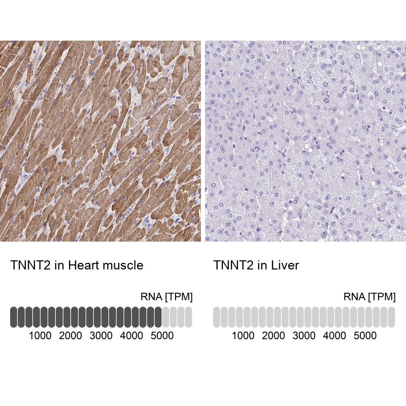
- Immunohistochemistry analysis in human heart muscle and liver tissues using HPA017888 antibody. Corresponding TNNT2 RNA-seq data are presented for the same tissues.
- Sample type
- Human
- Protocol
- Protocol