Explore
 Validate
 Learn
Learn Immunocytochemistry
 Immunohistochemistry
Antibody data
- Antibody Data
- Antigen structure
- References [1]
- Comments [0]
- Validations
- Immunocytochemistry [1]
Submit
Validation data
Reference
Comment
Report error
- Product number
- AMAb90961 - Provider product page

- Provider
- Atlas Antibodies
- Proper citation
- Atlas Antibodies Cat#AMAb90961, RRID:AB_2665734
- Product name
- Anti-NIFK
- Antibody type
- Monoclonal
- Description
- Monoclonal Antibody against Human NIFK, Clone ID: CL2240, Gene description: Nucleolar protein interacting with the fha domain of mki67, Alternative Gene Names: NIFK, hNIFK, Nopp34, Validated applications: IHC, ICC, Uniprot ID: Q9BYG3, Storage: Store at +4°C for short term storage. Long time storage is recommended at -20°C.
- Reactivity
- Human
- Host
- Mouse
- Conjugate
- Unconjugated
- Isotype
- IgG
- Antibody clone number
- CL2240
- Vial size
- 100 µl
- Concentration
- 1.0 mg/ml
- Storage
- Store at +4°C for short term storage. Long time storage is recommended at -20°C.
- Handling
- The antibody solution should be gently mixed before use.
Submitted references Dynamic chromosomal interactions and control of heterochromatin positioning by Ki-67.
van Schaik T, Manzo SG, Vouzas AE, Liu NQ, Teunissen H, de Wit E, Gilbert DM, van Steensel B
EMBO reports 2022 Dec 6;23(12):e55782
EMBO reports 2022 Dec 6;23(12):e55782
No comments: Submit comment
Supportive validation
- Submitted by
- Atlas Antibodies (provider)
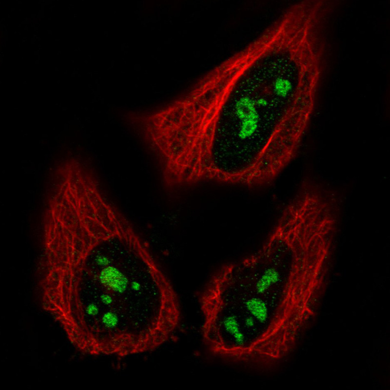
- Main image
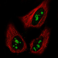
- Experimental details
- Immunofluorescence staining in HeLa cell line with Anti-MKI67IP monoclonal antibody, showing specific staining of nucleoli in green. Microtubule- and nuclear probes are visualized in red and blue respectively (where available).
- Sample type
- Human