Explore
 Validate
 Learn
Learn Western blot
 Immunocytochemistry
Antibody data
- Antibody Data
- Antigen structure
- References [8]
- Comments [0]
- Validations
- Immunocytochemistry [4]
- Flow cytometry [1]
- Other assay [4]
Submit
Validation data
Reference
Comment
Report error
- Product number
- PA1-077 - Provider product page

- Provider
- Invitrogen Antibodies
- Product name
- GM130 Polyclonal Antibody
- Antibody type
- Polyclonal
- Antigen
- Synthetic peptide
- Description
- PA1-077 detects GM130 from human samples. PA1-077 has been successfully used in Western blot, flow cytometry, and immunofluorescence procedures. By Western blot, PA1-077 detects a ~135 kDa band representing GM130 from Hela cell lysate. The PA1-077 immunogen is a synthetic peptide corresponding to residues G(368) Q V M E S V R Q L Q M E R D K(383) of human GM130. PA1-077 can be used with blocking peptide PEP-296.
- Reactivity
- Human
- Host
- Rabbit
- Isotype
- IgG
- Vial size
- 100 µg
- Concentration
- 1 mg/mL
- Storage
- -20° C, Avoid Freeze/Thaw Cycles
Submitted references Transcriptional and Post-Translational Regulation of Junctional Adhesion Molecule-B (JAM-B) in Leukocytes under Inflammatory Stimuli.
Exosome-mediated mRNA delivery in vivo is safe and can be used to induce SARS-CoV-2 immunity.
Overexpression of sortilin is associated with 5-FU resistance and poor prognosis in colorectal cancer.
TOM20-mediated transfer of Bcl2 from ER to MAM and mitochondria upon induction of apoptosis.
The Alzheimer's disease-associated C99 fragment of APP regulates cellular cholesterol trafficking.
A novel disorder involving dyshematopoiesis, inflammation, and HLH due to aberrant CDC42 function.
Human plasma C3 is essential for the development of memory B, but not T, lymphocytes.
Hyaluronan synthesis mediates the fibrotic response of keratocytes to transforming growth factor beta.
Day-Walsh PE, Keeble B, Pirabagar G, Fountain SJ, Kroon PA
International journal of molecular sciences 2022 Aug 3;23(15)
International journal of molecular sciences 2022 Aug 3;23(15)
Exosome-mediated mRNA delivery in vivo is safe and can be used to induce SARS-CoV-2 immunity.
Tsai SJ, Atai NA, Cacciottolo M, Nice J, Salehi A, Guo C, Sedgwick A, Kanagavelu S, Gould SJ
The Journal of biological chemistry 2021 Nov;297(5):101266
The Journal of biological chemistry 2021 Nov;297(5):101266
Overexpression of sortilin is associated with 5-FU resistance and poor prognosis in colorectal cancer.
Blondy S, Talbot H, Saada S, Christou N, Battu S, Pannequin J, Jauberteau MO, Lalloué F, Verdier M, Mathonnet M, Perraud A
Journal of cellular and molecular medicine 2021 Jan;25(1):47-60
Journal of cellular and molecular medicine 2021 Jan;25(1):47-60
TOM20-mediated transfer of Bcl2 from ER to MAM and mitochondria upon induction of apoptosis.
Lalier L, Mignard V, Joalland MP, Lanoé D, Cartron PF, Manon S, Vallette FM
Cell death & disease 2021 Feb 15;12(2):182
Cell death & disease 2021 Feb 15;12(2):182
The Alzheimer's disease-associated C99 fragment of APP regulates cellular cholesterol trafficking.
Montesinos J, Pera M, Larrea D, Guardia-Laguarta C, Agrawal RR, Velasco KR, Yun TD, Stavrovskaya IG, Xu Y, Koo SY, Snead AM, Sproul AA, Area-Gomez E
The EMBO journal 2020 Oct 15;39(20):e103791
The EMBO journal 2020 Oct 15;39(20):e103791
A novel disorder involving dyshematopoiesis, inflammation, and HLH due to aberrant CDC42 function.
Lam MT, Coppola S, Krumbach OHF, Prencipe G, Insalaco A, Cifaldi C, Brigida I, Zara E, Scala S, Di Cesare S, Martinelli S, Di Rocco M, Pascarella A, Niceta M, Pantaleoni F, Ciolfi A, Netter P, Carisey AF, Diehl M, Akbarzadeh M, Conti F, Merli P, Pastore A, Levi Mortera S, Camerini S, Farina L, Buchholzer M, Pannone L, Cao TN, Coban-Akdemir ZH, Jhangiani SN, Muzny DM, Gibbs RA, Basso-Ricci L, Chiriaco M, Dvorsky R, Putignani L, Carsetti R, Janning P, Stray-Pedersen A, Erichsen HC, Horne A, Bryceson YT, Torralba-Raga L, Ramme K, Rosti V, Bracaglia C, Messia V, Palma P, Finocchi A, Locatelli F, Chinn IK, Lupski JR, Mace EM, Cancrini C, Aiuti A, Ahmadian MR, Orange JS, De Benedetti F, Tartaglia M
The Journal of experimental medicine 2019 Dec 2;216(12):2778-2799
The Journal of experimental medicine 2019 Dec 2;216(12):2778-2799
Human plasma C3 is essential for the development of memory B, but not T, lymphocytes.
Jiménez-Reinoso A, Marin AV, Subias M, López-Lera A, Román-Ortiz E, Payne K, Ma CS, Arbore G, Kolev M, Freeley SJ, Kemper C, Tangye SG, Fernández-Malavé E, Rodríguez de Córdoba S, López-Trascasa M, Regueiro JR
The Journal of allergy and clinical immunology 2018 Mar;141(3):1151-1154.e14
The Journal of allergy and clinical immunology 2018 Mar;141(3):1151-1154.e14
Hyaluronan synthesis mediates the fibrotic response of keratocytes to transforming growth factor beta.
Guo N, Li X, Mann MM, Funderburgh ML, Du Y, Funderburgh JL
The Journal of biological chemistry 2010 Oct 15;285(42):32012-9
The Journal of biological chemistry 2010 Oct 15;285(42):32012-9
No comments: Submit comment
Supportive validation
- Submitted by
- Invitrogen Antibodies (provider)
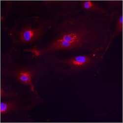
- Main image
- Experimental details
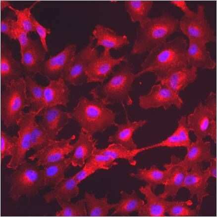
- Immunofluorescent analysis of GM130 using anti-GM130 polyclonal antibody (Product # PA1-077) shows staining in HMVEC Cells.
- Submitted by
- Invitrogen Antibodies (provider)
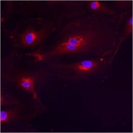
- Main image

- Experimental details
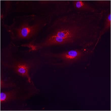
- Immunofluorescent analysis of GM130 using anti-GM130 polyclonal antibody (Product # PA1-077) shows staining in A549 Cells.
- Submitted by
- Invitrogen Antibodies (provider)
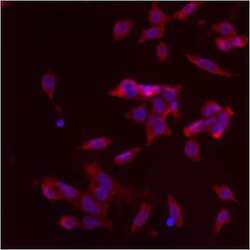
- Main image
- Experimental details
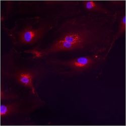
- Immunofluorescent analysis of GM130 using anti-GM130 polyclonal antibody (Product # PA1-077) shows staining in HMVEC Cells.
- Submitted by
- Invitrogen Antibodies (provider)
- Main image
- Experimental details
- Immunofluorescent analysis of GM130 using anti-GM130 polyclonal antibody (Product # PA1-077) shows staining in p19 Cells.

Supportive validation
- Submitted by
- Invitrogen Antibodies (provider)
- Main image
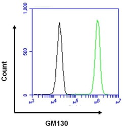
- Experimental details
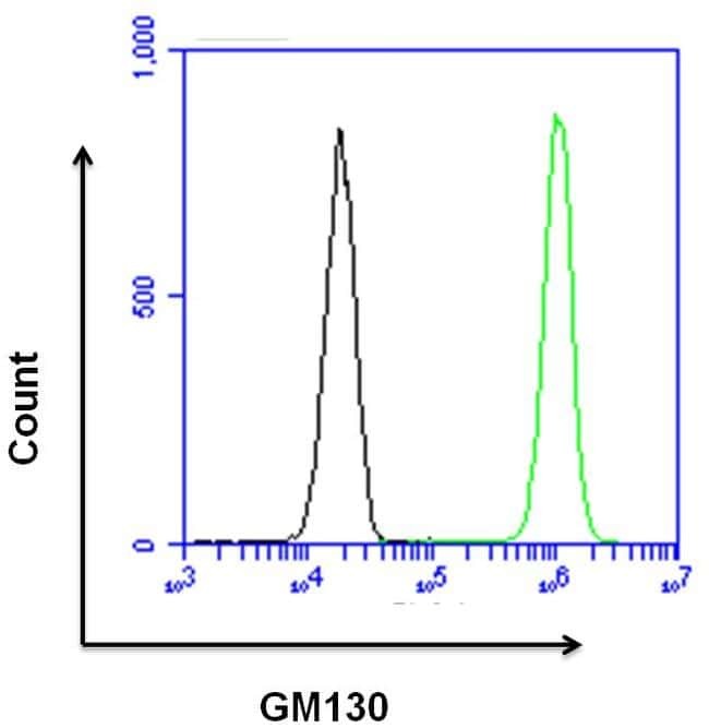
- Flow cytometry analysis of GM130 was done on HeLa cells. Cells were fixed, permeabilized and stained with a GM130 rabbit polyclonal antibody (Product # PA1-077, green histogram) or a rabbit IgG isotype control (Product # MA5-16384, black histogram) at a dilution of 10 µg/mL. After incubation for 1 hour on ice, the cells were labeled with a Goat anti-Rabbit IgG (H+L) Superclonal™ Secondary Antibody, Alexa Fluor® 647 conjugate (Product # A27040) at a dilution of 1:50 for 1 hour on ice. A representative 10,000 cells were acquired and analyzed for each sample.
Supportive validation
- Submitted by
- Invitrogen Antibodies (provider)
- Main image
- Experimental details
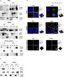
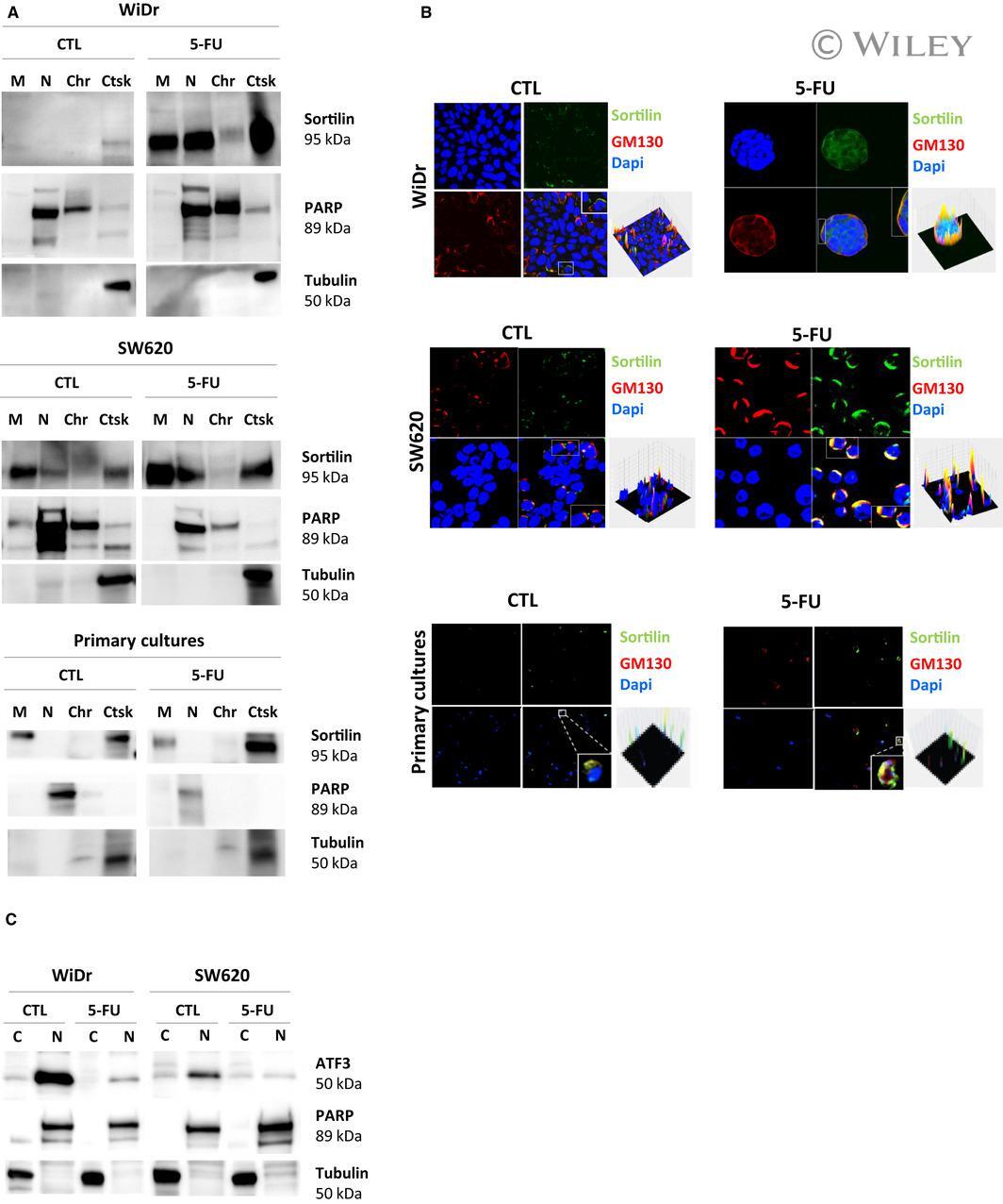
- 3 FIGURE Sortilin localization and transcriptional regulation upon long-term 5-FU treatment. A, To analyse sortilin localization, a subcellular fractionation was done on WiDr, SW620 and primary cultures treated by 5-FU. Extracted proteins from different cell component (C for cytoskeleton; M for membranes; N for nucleus; Chr for chromatin; Ctsk for cytosquelet) were subjected to Western blot analysis as described in materials and methods section. Anti-PARP antibody was used as a control for nuclear proteins, and anti-Tubulin antibody was used as a control of cytoplasmic proteins. B, Indirect immunofluorescence staining was analysed using confocal microscopy (LSM 510 META; Zeiss, Gottingen, Germany). Anti-GM130 was used as a Golgi marker and nuclei were stained with DAPI. Images were processed with the ZEN software application, and surface plots of the fluorescence data were generated with the image processing program ImageJ. C, ATF3 expression was analysed after subcellular fractionation of WiDr and SW620 as described just above
- Submitted by
- Invitrogen Antibodies (provider)
- Main image

- Experimental details
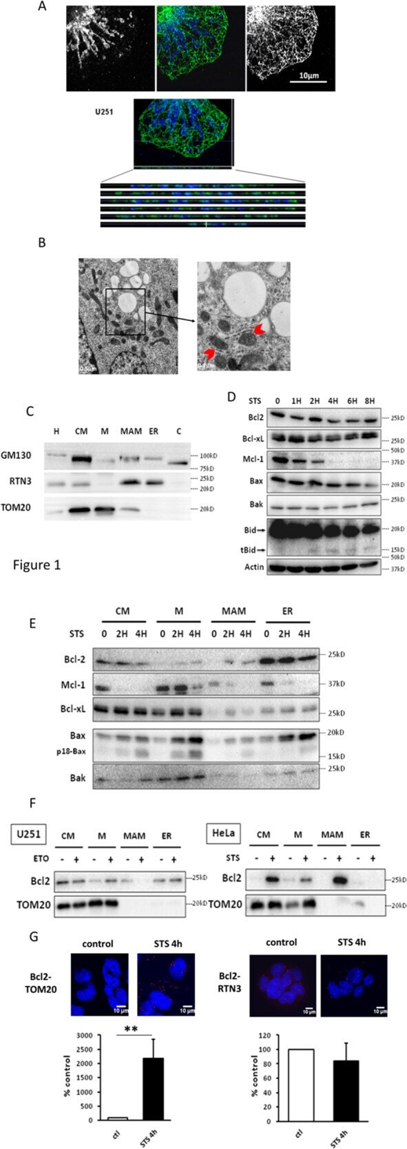
- Fig. 1 Bcl2 localization during the early phase of apoptosis. A Confocal analysis of MFN1 (blue) and RTN3 (green) shows the close proximity of mitochondria and ER in U251 cells. B Electron microscopy in U251 cells enables the visualization of MAM, formed by the juxtaposition of ER and mitochondria (red arrowheads). C Cell fractionation is realized in U251 cells and fractions are analyzed by western blot (GM130: Golgi protein, RTN3: ER protein, TOM20: mitochondria protein) (H: homogenate, CM: crude mitochondria, M: pure mitochondria, C: cytosol). The blots are representative of three independent fractionation experiments. D U251 cells were treated by STS and total lysates are analyzed by western blot. E U251 cells treated by STS for 0, 2 and 4 h are fractionated as in ( C ). Fractions are analyzed by western blot. F U251 cells were treated by etoposide for 24 h and HeLa cells by STS for 4 h. Cell fractionation was realized as in ( E ) and Bcl2 localization was analyzed by western blot. The blots shown in D - F are representative of three independent experiments. G Bcl2-TOM20 and Bcl2-RTN3 interactions were observed by PLA in U251 treated by STS.
- Submitted by
- Invitrogen Antibodies (provider)
- Main image
- Experimental details
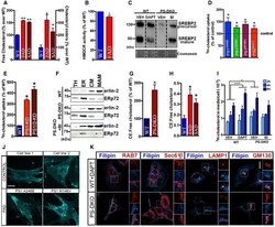
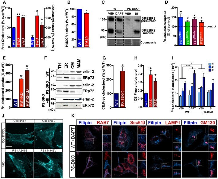
- Figure EV1 Increases in MAM -localized C99 induces cholesterol internalization and trafficking A Quantification of free cholesterol and cholesteryl ester levels in total homogenates from fibroblasts from familial (FAD) and sporadic (SAD) AD patients by lipidomics analysis. Lipid units are represented as molar mass over total moles of lipids analyzed (mol%). Graphs represent fold change over controls (WT). Unpaired t -test vs. WT ( n = 4-8; * P < 0.05, ** P < 0.01). B Quantification of HMGCR enzymatic activity showed a decreased rate of the de novo synthesis of cholesterol in fibroblasts from FAD patients. One-sample t -test vs. WT ( n = 3-4; * P < 0.05). C Measurement of SREBP2 levels by WB showed reductions in its mature/active form in DAPT-treated WT cells, and a reduction in both the full-length/precursor and mature forms in PS-DKO cells. Note how reductions in SREBP2 levels in mutant cells were abrogated upon BACE1 inhibition (BI). Coomassie staining is shown as a loading control. D, E Quantification of cholesterol uptake in (D) FAD fibroblasts and (E) neuroblastoma cell lines (Neuro-2a) where PS1, or both PS1 and PS2, had been transiently silenced. Dashed line indicates control levels. One-sample t -test vs. WT ( n = 3; * P < 0.05). F Western blot of isolated MAM and ER fractions used in Fig 1 E and F. Erlin-2 and ERp72 were used as MAM and ER markers, respectively (TH: total homogenate, CM: crude membrane). G, H Ratio of cholesteryl esters (CE):free cholesterol levels m
- Submitted by
- Invitrogen Antibodies (provider)
- Main image
- Experimental details
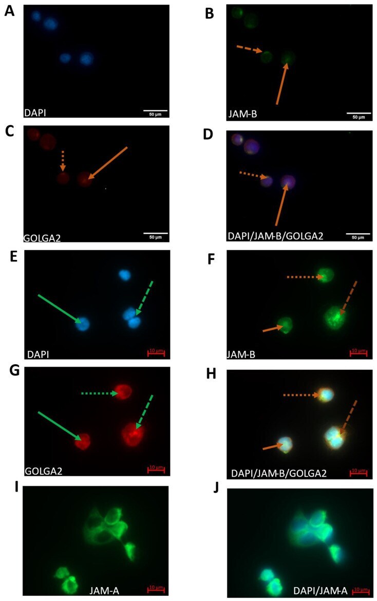
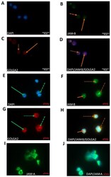
- Immunofluorescence of DAPI, JAM-B and G130 using a mouse anti-JAM-B monoclonal antibody (SC-293496) and an anti-GM130 polyclonal antibody (PA1-077) in THP-1 monocytes ( A - D ) and macrophages ( E - J ). ( A ) DAPI nuclear staining in THP-1 monocytes; ( B ) JAM-B staining in THP-1 monocytes; ( C ) G130 Golgi staining in THP-1 monocytes (stains peripheral membrane component of the cis-Golgi stack marker GOLGA2); ( D ) composite image showing co-localisation of JAM-B and G130; ( E ) DAPI staining in THP-1-differentiated macrophages; ( F ) JAM-B staining in THP-1-differentiated macrophages showing polarised staining (dotted line), in splitting nuclei (dashed line), inside the nucleus (solid line); ( G ) cis-Golgi stack marker GOLGA2; ( H ) composite image showing DAPI, JAM-B and GOLGA2 staining. At least three technical replicates were carried out. JAM-B staining contrasts with that of JAM-A ( I ) (JAM-A staining) and ( J ) (composite JAM-A and DAPI staining).