Explore
 Validate
 Learn
Learn Western blot
Antibody data
- Antibody Data
- Antigen structure
- References [0]
- Comments [0]
- Validations
- Western blot [4]
- Immunocytochemistry [1]
Submit
Validation data
Reference
Comment
Report error
- Product number
- PA1-32094 - Provider product page

- Provider
- Invitrogen Antibodies
- Product name
- Anti-KPNA2 Polyclonal Antibody
- Antibody type
- Polyclonal
- Antigen
- Synthetic peptide
- Description
- Recommended positive controls: The peptide used to generate this antibody is available for purchase (GTX26036-PEP).. Store product as a concentrated solution. Centrifuge briefly prior to opening the vial.
- Reactivity
- Human, Mouse, Rat
- Host
- Goat
- Isotype
- IgG
- Vial size
- 100 µg
- Concentration
- 0.5 mg/mL
- Storage
- Store at 4°C short term. For long term storage, store at -20°C, avoiding freeze/thaw cycles.
No comments: Submit comment
Supportive validation
- Submitted by
- Invitrogen Antibodies (provider)
- Main image

- Experimental details
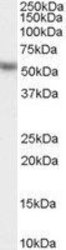
- Western blot analysis of KPNA2 in Hela lysate (RIPA buffer, 35 µg total protein per lane) using a KPNA2 polyclonal antibody (Product # PA1-32094) at a dilution of 0.1 µg/mL following incubation for 1 hour and detected using chemiluminescence.

- Submitted by
- Invitrogen Antibodies (provider)
- Main image
- Experimental details
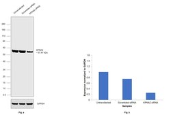
- Knockdown of KPNA2 was achieved by transfecting HEK-293 cells with KPNA2 specific siRNAs (Silencer® select Product # s7921, s7920). Western blot analysis (Fig. a) was performed using whole cell extracts from the KPNA2 knockdown cells (lane 3), non-specific scrambled siRNA transfected cells (lane 2) and untransfected cells (lane 1). The blots were probed using KPNA2 Polyclonal Antibody (Product # PA1-32094) and Rabbit anti-Goat IgG (H+L) Superclonal™ Recombinant Secondary Antibody, HRP (Product # A27014, 1:4000 dilution). Densitometric analysis of this western blot is shown in histogram (Fig. b). Decrease in signal upon siRNA mediated knock down confirms that antibody is specific to KPNA2.

- Submitted by
- Invitrogen Antibodies (provider)
- Main image

- Experimental details
- Western blot was performed using Anti-Importin subunit alpha-1 (KPNA2) Polyclonal Antibody (Product # PA1-32094) on whole cell extracts (30 µg lysate) of HEK-293 (Lane 1), A549 (Lane 2), HeLa (Lane 3), NIH/3T3 (Lane 4), PC-12 (Lane 5), LNCaP (Lane 6), Mouse Testis (Lane 7), Mouse Thymus (Lane 8), Mouse colon (Lane 9), Mouse Spleen (Lane 10) and Rat Testis (Lane 11). A 57.86 kDa band corresponding to Importin subunit alpha-1 (KPNA2) was observed except in Mouse colon which is reported to be negative. Resolved proteins were then transferred onto a nitrocellulose membrane (Product # IB23001) by iBlot® 2 Dry Blotting System (Product # IB21001). The blot was probed with the primary antibody (1:1000 dilution) and detected by Rabbit anti-Goat IgG (H+L) Superclonal™ Recombinant Secondary Antibody, HRP (Product # A27014, 1:4000 dilution) using the iBright FL 1000 (Product # A32752). Chemiluminescent detection was performed using Novex® ECL Chemiluminescent Substrate Reagent Kit (Product # WP20005).

- Submitted by
- Invitrogen Antibodies (provider)
- Main image
- Experimental details
- Western Blot analysis of KPNA2 was performed by loading 35 µg (in RIPA buffer) of HeLa lysates. Proteins were transferred to a membrane and probed with a KPNA2 Polyclonal Antibody (Product # PA1-32094) at a dilution of 0.03 µg/mL.

Supportive validation
- Submitted by
- Invitrogen Antibodies (provider)
- Main image

- Experimental details
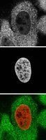
- Immunocytochemistry-Immunofluorescence analysis of KPNA2 in PFA fixed HeLa cells using KPNA2 Polyclonal Antibody (Product # PA1-32094) (Green). Red : HistonH2B-GFP fusion as DNA marker.