Explore
 Validate
 Learn
Learn Western blot
Antibody data
- Antibody Data
- Antigen structure
- References [0]
- Comments [0]
- Validations
- Western blot [2]
- Immunocytochemistry [1]
Submit
Validation data
Reference
Comment
Report error
- Product number
- MA5-17016 - Provider product page

- Provider
- Invitrogen Antibodies
- Product name
- KPNA2 Monoclonal Antibody (2G7)
- Antibody type
- Monoclonal
- Antigen
- Other
- Description
- MA5-17016 reacts with karyopherin 2 in human samples.
- Antibody clone number
- 2G7
- Concentration
- 1 mg/mL
No comments: Submit comment
Supportive validation
- Submitted by
- Invitrogen Antibodies (provider)
- Main image

- Experimental details
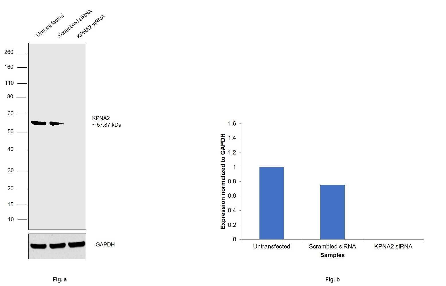
- Knockdown of KPNA2 was achieved by transfecting HEK-293 cells with KPNA2 specific siRNAs (Silencer® select Product # s7921, s7920). Western blot analysis (Fig. a) was performed using whole cell extracts from the KPNA2 knockdown cells (lane 3), non-specific scrambled siRNA transfected cells (lane 2) and untransfected cells (lane 1). The blots were probed using KPNA2 Monoclonal Antibody (Product # MA5-17016) and by F(ab)2-Rabbit anti-Rat IgG (H+L) Secondary Antibody, HRP (Product # PA1-29927, 1:4000 dilution). Densitometric analysis of this western blot is shown in histogram (Fig. b). Decrease in signal upon siRNA mediated knock down confirms that antibody is specific to KPNA2.
- Submitted by
- Invitrogen Antibodies (provider)
- Main image
- Experimental details
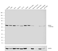
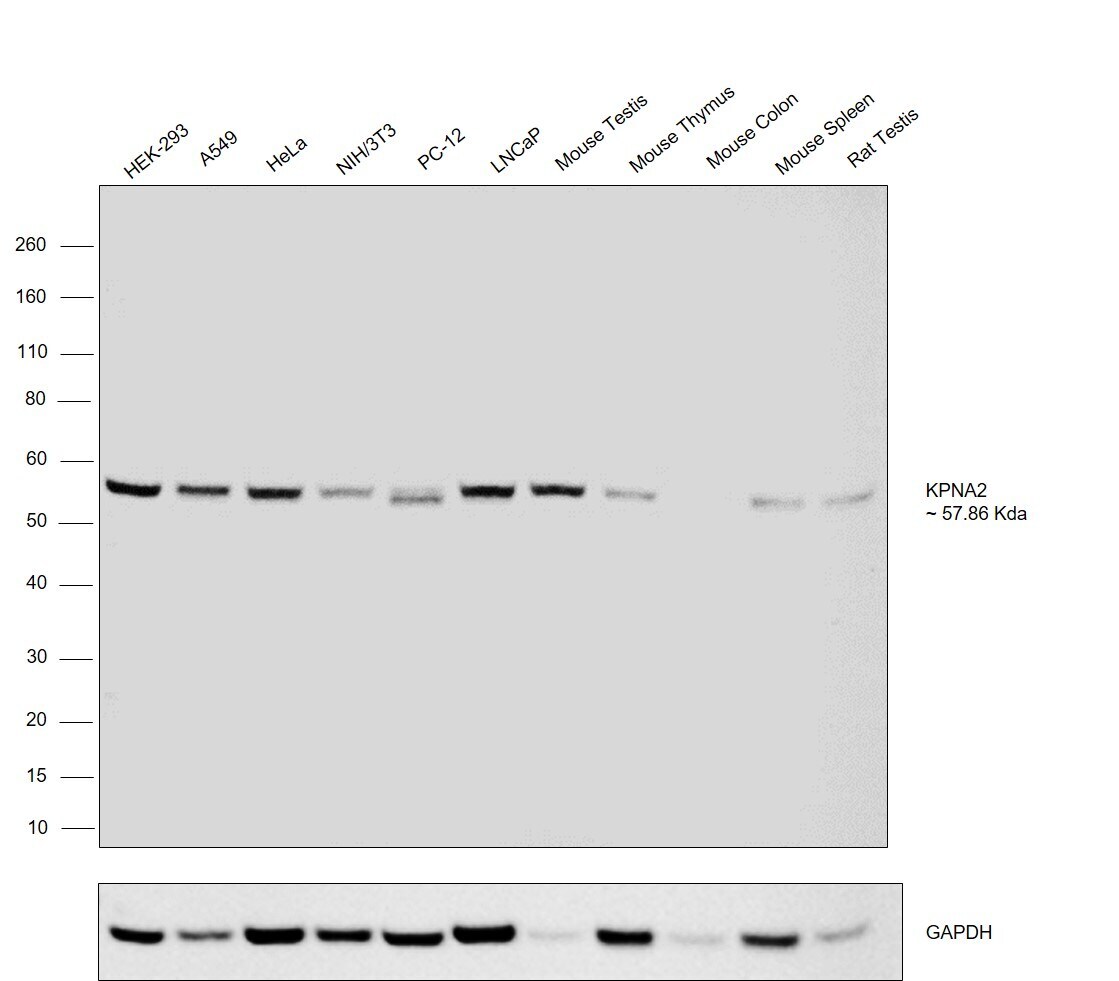
- Western blot was performed using Anti-Importin subunit alpha-1 (KPNA2) Monoclonal Antibody (Product # MA5-17016) on whole cell extracts (30 µg lysate) of HEK-293 (Lane 1), A549 (Lane 2), HeLa (Lane 3), NIH/3T3 (Lane 4), PC-12 (Lane 5), LNCaP (Lane 6), Mouse Testis (Lane 7), Mouse Thymus (Lane 8), Mouse colon (Lane 9), Mouse Spleen (Lane 10) and Rat Testis (Lane 11). A 57.86 kDa band corresponding to Importin subunit alpha-1 (KPNA2) was observed except in Mouse colon which is reported to be negative. Resolved proteins were then transferred onto a nitrocellulose membrane (Product # IB23001) by iBlot® 2 Dry Blotting System (Product # IB21001). The blot was probed with the primary antibody (1:1000 dilution) and detected by F(ab)2-Rabbit anti-Rat IgG (H+L) Secondary Antibody, HRP (Product # PA1-29927, 1:4000 dilution) using the iBright FL 1000 (Product # A32752). Chemiluminescent detection was performed using Novex® ECL Chemiluminescent Substrate Reagent Kit (Product # WP20005).
Supportive validation
- Submitted by
- Invitrogen Antibodies (provider)
- Main image

- Experimental details
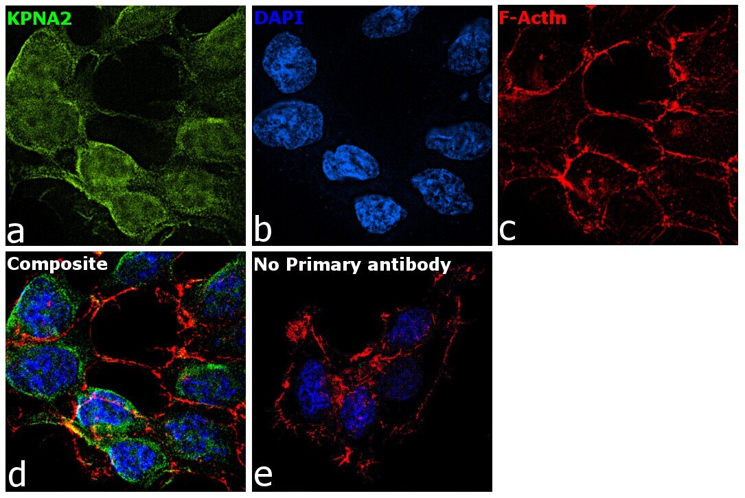
- Immunofluorescence analysis of KPNA2 was performed using HEK-293 cells. The cells were fixed with 4% paraformaldehyde for 10 minutes, permeabilized with 0.1% Triton™ X-100 for 15 minutes, and blocked with 2% BSA for 1 hour at room temperature. The cells were labeled with KPNA2 Monoclonal Antibody (Product # MA5-17016) at 1:100 dilution in 0.1% BSA and incubated overnight at 4 degree and then labeled with Goat anti-Rat IgG (H+L) Cross-Adsorbed Secondary Antibody, Alexa Fluor 488 (Product # A-11006) at a dilution of 1:2000 for 45 minutes at room temperature (Panel a: green). Nuclei (Panel b: blue) were stained with ProLong™ Diamond Antifade Mountant with DAPI (Product # P36962). F-actin (Panel c: red) was stained with Rhodamine Phalloidin (Product # R415, 1:300). Panel d represents the composite image showing cytoplasmic localization. Panel e represents control cells with no primary antibody to assess background. The images were captured at 60X magnification.