Explore
 Validate
 Learn
Learn Western blot
 Immunocytochemistry
Antibody data
- Antibody Data
- Antigen structure
- References [3]
- Comments [0]
- Validations
- Western blot [2]
- Immunohistochemistry [2]
- Flow cytometry [2]
Submit
Validation data
Reference
Comment
Report error
- Product number
- NBP2-31106 - Provider product page

- Provider
- Novus Biologicals
- Product name
- Mouse Monoclonal CD47 Antibody
- Antibody type
- Monoclonal
- Description
- Protein G purified.
- Reactivity
- Human, Mouse
- Host
- Mouse
- Isotype
- IgG
- Vial size
- 0.1 mg
- Concentration
- 1.0 mg/ml
- Storage
- Store at -20C. Avoid freeze-thaw cycles.
Submitted references Glutaminyl cyclase is an enzymatic modifier of the CD47- SIRPα axis and a target for cancer immunotherapy.
Surface-Enhanced Raman Scattering Nanoparticles for Multiplexed Imaging of Bladder Cancer Tissue Permeability and Molecular Phenotype.
CD47-blocking antibodies restore phagocytosis and prevent atherosclerosis.
Logtenberg MEW, Jansen JHM, Raaben M, Toebes M, Franke K, Brandsma AM, Matlung HL, Fauster A, Gomez-Eerland R, Bakker NAM, van der Schot S, Marijt KA, Verdoes M, Haanen JBAG, van den Berg JH, Neefjes J, van den Berg TK, Brummelkamp TR, Leusen JHW, Scheeren FA, Schumacher TN
Nature medicine 2019 Apr;25(4):612-619
Nature medicine 2019 Apr;25(4):612-619
Surface-Enhanced Raman Scattering Nanoparticles for Multiplexed Imaging of Bladder Cancer Tissue Permeability and Molecular Phenotype.
Davis RM, Kiss B, Trivedi DR, Metzner TJ, Liao JC, Gambhir SS
ACS nano 2018 Oct 23;12(10):9669-9679
ACS nano 2018 Oct 23;12(10):9669-9679
CD47-blocking antibodies restore phagocytosis and prevent atherosclerosis.
Kojima Y, Volkmer JP, McKenna K, Civelek M, Lusis AJ, Miller CL, Direnzo D, Nanda V, Ye J, Connolly AJ, Schadt EE, Quertermous T, Betancur P, Maegdefessel L, Matic LP, Hedin U, Weissman IL, Leeper NJ
Nature 2016 Aug 4;536(7614):86-90
Nature 2016 Aug 4;536(7614):86-90
No comments: Submit comment
Supportive validation
- Submitted by
- Novus Biologicals (provider)
- Main image
- Experimental details
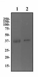
- Western Blot: CD47 Antibody (B6H12.2) [Azide Free] [NBP2-31106] - Human brain (lane 1) and testis (lane 2) protein was separated on a 12% gel by SDS-PAGE. Protein was transferred to PVDF membrane, blocked and then probed with 2 ug/ml of anti-CD47. CD47 protein was detected using an anti-mouse HRP secondary antibody.

- Submitted by
- Novus Biologicals (provider)
- Main image
- Experimental details
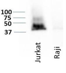
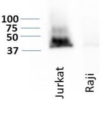
- Western Blot: CD47 Antibody (B6H12.2) - Azide Free [NBP2-31106] - Detection of CD47 expression on two hematological cancer cell lines, Jurkat and Raji. WB image submitted by a verified customer review.
Supportive validation
- Submitted by
- Novus Biologicals (provider)
- Main image

- Experimental details
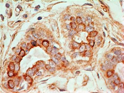
- Immunohistochemistry-Paraffin: CD47 Antibody (B6H12.2) - Azide Free [NBP2-31106] - Tissue section of human normal breast using mouse monoclonal CD47 antibody (clone B6H12.2) at 0.5ug/ml concentration. The ductal/acinar epithelial cells in the breast section developed specific membrane-cytoplasmic staining.
- Submitted by
- Novus Biologicals (provider)
- Main image
- Experimental details
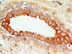
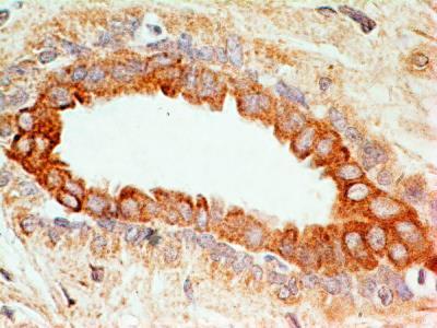
- Immunohistochemistry-Paraffin: CD47 Antibody (B6H12.2) - Azide Free [NBP2-31106] - Tissue section of human normal breast using mouse monoclonal CD47 antibody (clone B6H12.2) at 0.5ug/ml concentration. The ductal/acinar epithelial cells in the breast section developed specific membrane-cytoplasmic staining.
Supportive validation
- Submitted by
- Novus Biologicals (provider)
- Main image
- Experimental details
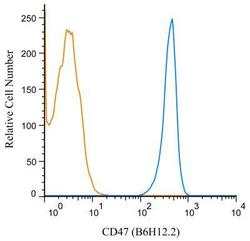
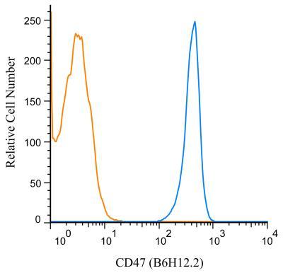
- Flow (Cell Surface): CD47 Antibody (B6H12.2) - Azide Free [NBP2-31106] - A surface stain was performed on human peripheral blood lymphocytes with CD47 (B6H12.2) antibody NBP2-31106 (blue) and a matched isotype control NBP2-27287 (orange). Cells were incubated in an antibody dilution of 1 ug/mL for 20 minutes at room temperature, followed by mouse F(ab)2 IgG (H+L) APC-conjugated secondary antibody [F0101B, R&D Systems].
- Submitted by
- Novus Biologicals (provider)
- Main image
- Experimental details
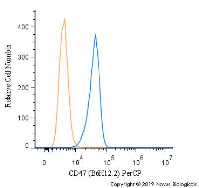
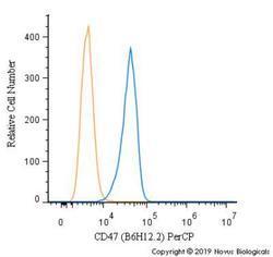
- Flow Cytometry: CD47 Antibody (B6H12.2) - Azide Free [NBP2-31106] - A surface stain was performed on A431 cells with CD47 Antibody [B6H12.2] NBP2-31106PCP (blue) and a matched isotype control (orange). Cells were incubated in an antibody dilution of 5 ug/mL for 20 minutes at room temperature. Both antibodies were conjugated to PerCP.