 Explore
 Validate
 Learn
Learn Western blot
 Flow cytometry
Antibody data
- Antibody Data
- Antigen structure
- References [6]
- Comments [0]
- Validations
- Flow cytometry [1]
- Other assay [2]
Submit
Validation data
Reference
Comment
Report error
- Product number
- 14-0478-82 - Provider product page

- Provider
- Invitrogen Antibodies
- Product name
- CD47 Monoclonal Antibody (2D3), eBioscience™
- Antibody type
- Monoclonal
- Antigen
- Other
- Description
- Description: The monoclonal antibody 2D3 reacts to CD47 also known as integrin-associated protein (IAP), and neurophilin. CD47 is a glycosylated five transmembrane protein with a small alternatively spliced cytoplasmic domain. CD47 is involved in adhesion through interactions with SIRP (signal regulator protein) and is non-covalently associated with beta3 integrins CD51/CD61 and CD41/CD61. Furthermore this interaction can mediate bi-directional signaling to modify neural synaptic activity and regulate the phagocytic activities of macrophages. CD47 is the receptor for thrombospondin. T cell expression of CD47 can mediate activation or apoptosis (in the presence of high levels of thrombospondin). Expression is found in the majority of hematopoietic cells including T and B cells, monocytes, platelets and erythrocytes (as part of the Rh complex). Expression is also found in non-hematopoietic cells. Applications Reported: This 2D3 antibody has been reported for use in flow cytometric analysis, and immunoblotting (WB) under nonreducing conditions. Applications Tested: This 2D3 antibody has been tested by flow cytometric analysis of normal human peripheral blood cells. This can be used at less than or equal to 0.5 µg per test. A test is defined as the amount (µg) of antibody that will stain a cell sample in a final volume of 100 µL. Cell number should be determined empirically but can range from 10^5 to 10^8 cells/test. It is recommended that the antibody be carefully titrated for optimal performance in the assay of interest. Purity: Greater than 90%, as determined by SDS-PAGE. Aggregation: Less than 10%, as determined by HPLC. Filtration: 0.2 µm post-manufacturing filtered.
- Reactivity
- Human
- Host
- Mouse
- Isotype
- IgG
- Antibody clone number
- 2D3
- Vial size
- 100 µg
- Concentration
- 0.5 mg/mL
- Storage
- 4° C
Submitted references Macrophages show higher levels of engulfment after disruption of cis interactions between CD47 and the checkpoint receptor SIRPα.
Accelerated Wound Healing by Fibroblasts Differentiated from Human Embryonic Stem Cell-Derived Mesenchymal Stem Cells in a Pressure Ulcer Animal Model.
CD47 agonist peptides induce programmed cell death in refractory chronic lymphocytic leukemia B cells via PLCγ1 activation: evidence from mice and humans.
Novel CD47: SIRPα dependent mechanism for the activation of STAT3 in antigen-presenting cell.
Regulation of cross-linked actin network (CLAN) formation in human trabecular meshwork (HTM) cells by convergence of distinct beta1 and beta3 integrin pathways.
Species- and cell type-specific interactions between CD47 and human SIRPalpha.
Hayes BH, Tsai RK, Dooling LJ, Kadu S, Lee JY, Pantano D, Rodriguez PL, Subramanian S, Shin JW, Discher DE
Journal of cell science 2020 Mar 6;133(5)
Journal of cell science 2020 Mar 6;133(5)
Accelerated Wound Healing by Fibroblasts Differentiated from Human Embryonic Stem Cell-Derived Mesenchymal Stem Cells in a Pressure Ulcer Animal Model.
Yoon D, Yoon D, Sim H, Hwang I, Lee JS, Chun W
Stem cells international 2018;2018:4789568
Stem cells international 2018;2018:4789568
CD47 agonist peptides induce programmed cell death in refractory chronic lymphocytic leukemia B cells via PLCγ1 activation: evidence from mice and humans.
Martinez-Torres AC, Quiney C, Attout T, Boullet H, Herbi L, Vela L, Barbier S, Chateau D, Chapiro E, Nguyen-Khac F, Davi F, Le Garff-Tavernier M, Moumné R, Sarfati M, Karoyan P, Merle-Béral H, Launay P, Susin SA
PLoS medicine 2015 Mar;12(3):e1001796
PLoS medicine 2015 Mar;12(3):e1001796
Novel CD47: SIRPα dependent mechanism for the activation of STAT3 in antigen-presenting cell.
Toledano N, Gur-Wahnon D, Ben-Yehuda A, Rachmilewitz J
PloS one 2013;8(9):e75595
PloS one 2013;8(9):e75595
Regulation of cross-linked actin network (CLAN) formation in human trabecular meshwork (HTM) cells by convergence of distinct beta1 and beta3 integrin pathways.
Filla MS, Schwinn MK, Sheibani N, Kaufman PL, Peters DM
Investigative ophthalmology & visual science 2009 Dec;50(12):5723-31
Investigative ophthalmology & visual science 2009 Dec;50(12):5723-31
Species- and cell type-specific interactions between CD47 and human SIRPalpha.
Subramanian S, Parthasarathy R, Sen S, Boder ET, Discher DE
Blood 2006 Mar 15;107(6):2548-56
Blood 2006 Mar 15;107(6):2548-56
No comments: Submit comment
Supportive validation
- Submitted by
- Invitrogen Antibodies (provider)
- Main image
- Experimental details
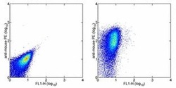
- Staining of normal human peripheral blood cells with 0.25 µg of Mouse IgG1 kappa Isotype Control Purified (Product # 14-4714-82) (left) or 0.25 µg of Purified anti-human CD47 Purified (right) followed by Anti-Mouse IgG PE (Product # 12-4012). Total lymphocytes were used for analysis.

Supportive validation
- Submitted by
- Invitrogen Antibodies (provider)
- Main image

- Experimental details
- NULL

- Submitted by
- Invitrogen Antibodies (provider)
- Main image
- Experimental details
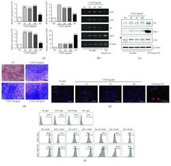
- Figure 1 Fibrogenic differentiation of human embryonic stem cell-derived mesenchymal stem cells (hESC-MSCs) upon stimulation with connective tissue growth factor (CTGF). hESC-MSCs were differentiated into fibroblasts by treatment with various concentrations of connective tissue growth factor (CTGF) for 4 weeks. Normal skin fibroblasts (Detroit 551) were also used as a positive control. (a) mRNA levels of fibroblast-related genes in hESC-MSCs after CTGF treatment were determined by the real-time polymerase chain reaction (PCR) ( n = 3, one-way ANOVA; ** p < 0.01 and **** p < 0.0001). (b) Collagen (Col)1, Col3, fibronectin (FN), and fibroblast-specific protein- (FSP-) 1 mRNA levels were determined by PCR. (c) FN, FSP-1, Col1, and beta -actin protein levels in hESC-MSCs following CTGF treatment were determined by immunoblotting. (d) Masson's trichrome was used to detect collagen fibers. (e) hESC-MSCs were immunostained to detect collagen I (Col1) following CTGF treatment. 4',6'-Diamidino-2-phenylindole (DAPI) was used for nuclear counterstaining. (f) Flow cytometry analysis of hESC-MSCs. After expansion of hESC-MSCs and hESC-MSC-Fbs, cells were trypsinized and stained with specific markers for CD29, CD47, CD73, CD90, CD91, CD105, and CD166.
